 Movie
Movie Controller


+ Open data

- Basic information
| Entry | Database: PDB / ID: 2j61 | ||||||
|---|---|---|---|---|---|---|---|
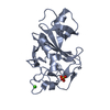
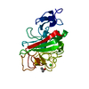
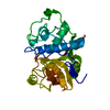
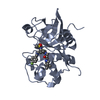
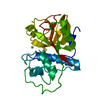
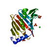
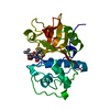
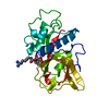
| Title | L-ficolin complexed to N-acetylglucosamine (forme C) | ||||||
 Components Components | (FICOLIN-2) x 2 | ||||||
 Keywords Keywords | LECTIN / GLYCOPROTEIN / FICRINOGEN-LIKE / INNATE IMMUNITY / PATTERN RECOGNITION PROTEIN / COLLAGEN / IMMUNOLOGY / LECTIN- LIKE | ||||||
| Function / homology |  Function and homology information Function and homology informationlipoteichoic acid immune receptor activity / mannan binding / recognition of apoptotic cell / Ficolins bind to repetitive carbohydrate structures on the target cell surface / Lectin pathway of complement activation / positive regulation of opsonization / complement activation, lectin pathway / opsonization / cell surface pattern recognition receptor signaling pathway / collagen trimer ...lipoteichoic acid immune receptor activity / mannan binding / recognition of apoptotic cell / Ficolins bind to repetitive carbohydrate structures on the target cell surface / Lectin pathway of complement activation / positive regulation of opsonization / complement activation, lectin pathway / opsonization / cell surface pattern recognition receptor signaling pathway / collagen trimer / carbohydrate derivative binding / symbiont cell surface / serine-type endopeptidase complex / proteoglycan binding / Initial triggering of complement / pattern recognition receptor activity / antigen binding / calcium-dependent protein binding / blood microparticle / defense response to Gram-negative bacterium / defense response to Gram-positive bacterium / signaling receptor binding / external side of plasma membrane / proteolysis / : / extracellular exosome / extracellular region / metal ion binding Similarity search - Function | ||||||
| Biological species |  HOMO SAPIENS (human) HOMO SAPIENS (human) | ||||||
| Method |  X-RAY DIFFRACTION / SYNCHROTRON / MOLECULAR REPLACEMENT / Resolution: 2.7 Å X-RAY DIFFRACTION / SYNCHROTRON / MOLECULAR REPLACEMENT / Resolution: 2.7 Å | ||||||
 Authors Authors | Garlatti, V. / Gaboriaud, C. | ||||||
 Citation Citation | Journal: Embo J. / Year: 2007 Title: Structural Insights Into the Innate Immune Recognition Specificities of L- and H-Ficolins. Authors: Garlatti, V. / Belloy, N. / Martin, L. / Lacroix, M. / Matsushita, M. / Endo, Y. / Fujita, T. / Fontecilla-Camps, J.C. / Arlaud, G.J. / Thielens, N.M. / Gaboriaud, C. | ||||||
| History |
| ||||||
| Remark 700 | SHEET THE SHEET STRUCTURE OF THIS MOLECULE IS BIFURCATED. IN ORDER TO REPRESENT THIS FEATURE IN ... SHEET THE SHEET STRUCTURE OF THIS MOLECULE IS BIFURCATED. IN ORDER TO REPRESENT THIS FEATURE IN THE SHEET RECORDS BELOW, TWO SHEETS ARE DEFINED. |
- Structure visualization
| Structure viewer | Molecule: MolmilJmol/JSmol |
|---|
- Downloads & links
-Download
| PDBx/mmCIF format | 2j61.cif.gz | 102.5 KB | Display | PDBx/mmCIF format |
|---|---|---|---|---|
| PDB format | pdb2j61.ent.gz | 78.7 KB | Display | PDB format |
| PDBx/mmJSON format | 2j61.json.gz | Tree view | PDBx/mmJSON format | |
| Others |  Other downloads Other downloads |
-Validation report
| Arichive directory | https://data.pdbj.org/pub/pdb/validation_reports/j6/2j61ftp://data.pdbj.org/pub/pdb/validation_reports/j6/2j61 | HTTPS FTP |
|---|
-Related structure data
| Related structure data |  2j0gC  2j0hC  2j0yC  2j1gC  2j2pC  2j3fC  2j3gC  2j3oC  2j3uC  2j5zC  2j60C  2j64C 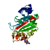 1jc9S C: citing same article ( S: Starting model for refinement |
|---|---|
| Similar structure data |

-Links
 PDBj
PDBj


- Assembly
| Deposited unit | 
| ||||||||
|---|---|---|---|---|---|---|---|---|---|
| 1 | 
| ||||||||
| 2 | 
| ||||||||
| Unit cell |
| ||||||||
| Components on special symmetry positions |
|
-Components
| #1: Protein | Mass: 24757.332 Da / Num. of mol.: 1 / Fragment: BINDING DOMAIN, RESIDUES 97-313 Source method: isolated from a genetically manipulated source Source: (gene. exp.) HOMO SAPIENS (human) / Tissue: PLASMA / Cell line (production host): High Five / Production host:  TRICHOPLUSIA NI (cabbage looper) / References: UniProt: Q15485 TRICHOPLUSIA NI (cabbage looper) / References: UniProt: Q15485 | ||||||||
|---|---|---|---|---|---|---|---|---|---|
| #2: Protein | Mass: 24729.318 Da / Num. of mol.: 1 / Fragment: BINDING DOMAIN, RESIDUES 97-313 Source method: isolated from a genetically manipulated source Source: (gene. exp.) HOMO SAPIENS (human) / Tissue: PLASMA / Cell line (production host): High Five / Production host: TRICHOPLUSIA NI (cabbage looper) / References: UniProt: Q15485 | ||||||||
| #3: Chemical |   Mass: 40.078 Da / Num. of mol.: 2 / Source method: obtained synthetically / Formula: Ca Mass: 40.078 Da / Num. of mol.: 2 / Source method: obtained synthetically / Formula: Ca#4: Sugar | ChemComp-NAG / |   Type: D-saccharide, beta linking / Mass: 221.208 Da / Num. of mol.: 1 Type: D-saccharide, beta linking / Mass: 221.208 Da / Num. of mol.: 1Source method: isolated from a genetically manipulated source Formula: C8H15NO6 #5: Water | ChemComp-HOH / |  Mass: 18.015 Da / Num. of mol.: 62 / Source method: isolated from a natural source / Formula: H2O Mass: 18.015 Da / Num. of mol.: 62 / Source method: isolated from a natural source / Formula: H2OHas protein modification | Y | Sequence details | DOMAINE C TERMINAL DIFFERENCE BETWEEN UNP SEQUENCE AND THE PROTEIN CRYSTALLIZED BECAUSE IT EXISTS ...DOMAINE C TERMINAL DIFFERENCE | |
-Experimental details
-Experiment
| Experiment | Method: X-RAY DIFFRACTION |
|---|
- Sample preparation
| Crystal | Density Matthews: 3.16 Å3/Da / Density % sol: 60.8 % |
|---|
-Data collection
| Diffraction | Mean temperature: 100 K |
|---|---|
| Diffraction source | Source: SYNCHROTRON / Site: ESRF  / Beamline: BM30A / Wavelength: 0.9206 / Beamline: BM30A / Wavelength: 0.9206 |
| Detector | Type: MARRESEARCH / Detector: CCD / Date: Apr 4, 2004 |
| Radiation | Protocol: SINGLE WAVELENGTH / Monochromatic (M) / Laue (L): M / Scattering type: x-ray |
| Radiation wavelength | Wavelength: 0.9206 Å / Relative weight: 1 |
| Reflection | Resolution: 2.7→19.39 Å / Num. obs: 16911 / % possible obs: 99.8 % / Observed criterion σ(I): 0 / Redundancy: 6.9 % / Rmerge(I) obs: 0.05 / Net I/σ(I): 24.6 |
- Processing
| Software |
| ||||||||||||||||||||||||||||||||||||||||||||||||||||||||||||||||||||||||||||||||||||||||||||||||||||||||||||||||||||||||||||||||||||||||||||||||||||||||||||||||||||||||||||||||||||||
|---|---|---|---|---|---|---|---|---|---|---|---|---|---|---|---|---|---|---|---|---|---|---|---|---|---|---|---|---|---|---|---|---|---|---|---|---|---|---|---|---|---|---|---|---|---|---|---|---|---|---|---|---|---|---|---|---|---|---|---|---|---|---|---|---|---|---|---|---|---|---|---|---|---|---|---|---|---|---|---|---|---|---|---|---|---|---|---|---|---|---|---|---|---|---|---|---|---|---|---|---|---|---|---|---|---|---|---|---|---|---|---|---|---|---|---|---|---|---|---|---|---|---|---|---|---|---|---|---|---|---|---|---|---|---|---|---|---|---|---|---|---|---|---|---|---|---|---|---|---|---|---|---|---|---|---|---|---|---|---|---|---|---|---|---|---|---|---|---|---|---|---|---|---|---|---|---|---|---|---|---|---|---|---|
| Refinement | Method to determine structure: MOLECULAR REPLACEMENT Starting model: PDB ENTRY 1JC9 Resolution: 2.7→15 Å / Cor.coef. Fo:Fc: 0.928 / Cor.coef. Fo:Fc free: 0.867 / SU B: 16.011 / SU ML: 0.331 / Cross valid method: THROUGHOUT / ESU R: 0.843 / ESU R Free: 0.38 / Stereochemistry target values: MAXIMUM LIKELIHOOD / Details: HYDROGENS HAVE BEEN ADDED IN THE RIDING POSITIONS.
| ||||||||||||||||||||||||||||||||||||||||||||||||||||||||||||||||||||||||||||||||||||||||||||||||||||||||||||||||||||||||||||||||||||||||||||||||||||||||||||||||||||||||||||||||||||||
| Solvent computation | Ion probe radii: 0.8 Å / Shrinkage radii: 0.8 Å / VDW probe radii: 1.4 Å / Solvent model: BABINET MODEL WITH MASK | ||||||||||||||||||||||||||||||||||||||||||||||||||||||||||||||||||||||||||||||||||||||||||||||||||||||||||||||||||||||||||||||||||||||||||||||||||||||||||||||||||||||||||||||||||||||
| Displacement parameters | Biso mean: 56.49 Å2
| ||||||||||||||||||||||||||||||||||||||||||||||||||||||||||||||||||||||||||||||||||||||||||||||||||||||||||||||||||||||||||||||||||||||||||||||||||||||||||||||||||||||||||||||||||||||
| Refinement step | Cycle: LAST / Resolution: 2.7→15 Å
| ||||||||||||||||||||||||||||||||||||||||||||||||||||||||||||||||||||||||||||||||||||||||||||||||||||||||||||||||||||||||||||||||||||||||||||||||||||||||||||||||||||||||||||||||||||||
| Refine LS restraints |
|