 Movie
Movie Controller

[English] 日本語
 Yorodumi
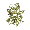
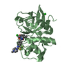
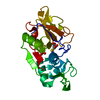
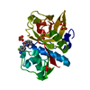
- PDB-1meg: CRYSTAL STRUCTURE OF A CARICAIN D158E MUTANT IN COMPLEX WITH E-64 -
+ Open data

- Basic information
| Entry | Database: PDB / ID: 1meg | ||||||
|---|---|---|---|---|---|---|---|
| Title | CRYSTAL STRUCTURE OF A CARICAIN D158E MUTANT IN COMPLEX WITH E-64 | ||||||
 Components Components | CARICAIN | ||||||
 Keywords Keywords | HYDROLASE / CYSTEINE PROTEINASE / THIOL PROTEASE | ||||||
| Function / homology |  Function and homology information Function and homology information | ||||||
| Biological species |  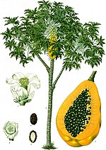 Carica papaya (papaya) Carica papaya (papaya) | ||||||
| Method |  X-RAY DIFFRACTION / SYNCHROTRON / Resolution: 2 Å X-RAY DIFFRACTION / SYNCHROTRON / Resolution: 2 Å | ||||||
 Authors Authors | Katerelos, N.A. | ||||||
 Citation Citation | Journal: FEBS Lett. / Year: 1996 Title: Crystal structure of a caricain D158E mutant in complex with E-64. Authors: Katerelos, N.A. / Taylor, M.A. / Scott, M. / Goodenough, P.W. / Pickersgill, R.W. #1: Journal: To be PublishedTitle: Rapid Kinetics Studies and Structural Determination of a Cysteine Proteinase Mutant Implies that Residue Asp 158 in Caricain Has a Major Effect Upon the Ability of the Active Site Histidine to ...Title: Rapid Kinetics Studies and Structural Determination of a Cysteine Proteinase Mutant Implies that Residue Asp 158 in Caricain Has a Major Effect Upon the Ability of the Active Site Histidine to Protonate a Dipyridyl Probe Authors: Katerelos, A. / Goodenough, P.W. #2: Journal: Protein Eng. / Year: 1994Title: An Unequivocal Example of Cysteine Proteinase Activity Affected by Multiple Electrostatic Interactions Authors: Taylor, M.A. / Baker, K.C. / Connerton, I.F. / Cummings, N.J. / Harris, G.W. / Henderson, I.M. / Jones, S.T. / Pickersgill, R.W. / Sumner, I.G. / Warwicker, J. / al., et #3: Journal: Gene / Year: 1993Title: Nucleotide Sequence and Expression in Escherichia Coli of Cdnas Encoding Papaya Proteinase Omega from Carica Papaya Authors: Revell, D.F. / Cummings, N.J. / Baker, K.C. / Collins, M.E. / Taylor, M.A. / Sumner, I.G. / Pickersgill, R.W. / Connerton, I.F. / Goodenough, P.W. #4: Journal: Protein Eng. / Year: 1992Title: Active Papain Renatured and Processed from Insoluble Recombinant Propapain Expressed in Escherichia Coli Authors: Taylor, M.A. / Pratt, K.A. / Revell, D.F. / Baker, K.C. / Sumner, I.G. / Goodenough, P.W. | ||||||
| History |
|
- Structure visualization
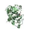
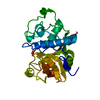
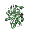
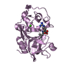
| Structure viewer | Molecule: MolmilJmol/JSmol |
|---|
- Downloads & links
-Download
| PDBx/mmCIF format | 1meg.cif.gz | 56 KB | Display | PDBx/mmCIF format |
|---|---|---|---|---|
| PDB format | pdb1meg.ent.gz | 39.8 KB | Display | PDB format |
| PDBx/mmJSON format | 1meg.json.gz | Tree view | PDBx/mmJSON format | |
| Others |  Other downloads Other downloads |
-Validation report
| Arichive directory | https://data.pdbj.org/pub/pdb/validation_reports/me/1megftp://data.pdbj.org/pub/pdb/validation_reports/me/1meg | HTTPS FTP |
|---|
-Related structure data
| Similar structure data |
|---|


-Links
 PDBj
PDBj


- Assembly
| Deposited unit | 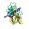
| ||||||||
|---|---|---|---|---|---|---|---|---|---|
| 1 |
| ||||||||
| Unit cell |
|
-Components
| #1: Protein | Mass: 23339.770 Da / Num. of mol.: 1 / Mutation: D158E Source method: isolated from a genetically manipulated source Source: (gene. exp.) Carica papaya (papaya) / Plasmid: PLYSS / Production host:  |
|---|---|
| #2: Chemical | ChemComp-E64 /   Mass: 360.429 Da / Num. of mol.: 1 / Source method: obtained synthetically / Formula: C15H30N5O5 Mass: 360.429 Da / Num. of mol.: 1 / Source method: obtained synthetically / Formula: C15H30N5O5 |
| #3: Chemical | ChemComp-EOH /   Mass: 46.068 Da / Num. of mol.: 1 / Source method: obtained synthetically / Formula: C2H6O Mass: 46.068 Da / Num. of mol.: 1 / Source method: obtained synthetically / Formula: C2H6O |
| #4: Water | ChemComp-HOH /  Mass: 18.015 Da / Num. of mol.: 94 / Source method: isolated from a natural source / Formula: H2O Mass: 18.015 Da / Num. of mol.: 94 / Source method: isolated from a natural source / Formula: H2O |
| Has protein modification | Y |
-Experimental details
-Experiment
| Experiment | Method: X-RAY DIFFRACTION |
|---|
- Sample preparation
| Crystal | Density Matthews: 2.24 Å3/Da / Density % sol: 34.2 % | ||||||||||||||||||||||||||||||
|---|---|---|---|---|---|---|---|---|---|---|---|---|---|---|---|---|---|---|---|---|---|---|---|---|---|---|---|---|---|---|---|
| Crystal | *PLUS | ||||||||||||||||||||||||||||||
| Crystal grow | *PLUS Temperature: 18 ℃ / pH: 8 / Method: vapor diffusion, hanging drop | ||||||||||||||||||||||||||||||
| Components of the solutions | *PLUS
|
-Data collection
| Diffraction source | Source: SYNCHROTRON / Site: Photon Factory  / Beamline: BL-6A / Wavelength: 1 / Beamline: BL-6A / Wavelength: 1 |
|---|---|
| Detector | Type: FUJI / Detector: IMAGE PLATE / Date: Nov 10, 1994 |
| Radiation | Monochromatic (M) / Laue (L): M / Scattering type: x-ray |
| Radiation wavelength | Wavelength: 1 Å / Relative weight: 1 |
| Reflection | Num. obs: 12010 / % possible obs: 86 % / Redundancy: 3.5 % / Rmerge(I) obs: 0.06 |
| Reflection | *PLUS Highest resolution: 2 Å / Lowest resolution: 7.72 Å / % possible obs: 86 % / Num. measured all: 42446 / Rmerge(I) obs: 0.06 |
- Processing
| Software |
| ||||||||||||||||||||||||||||||||||||||||||||||||||||||||||||
|---|---|---|---|---|---|---|---|---|---|---|---|---|---|---|---|---|---|---|---|---|---|---|---|---|---|---|---|---|---|---|---|---|---|---|---|---|---|---|---|---|---|---|---|---|---|---|---|---|---|---|---|---|---|---|---|---|---|---|---|---|---|
| Refinement | Resolution: 2→10 Å / σ(F): 2 Details: ATOMS C13, C14, C15, N3, C16, N4 AND N5 OF E64 WERE NOT VISIBLE IN DENSITY BECAUSE OF THE HIGH MOBILITY OF THAT PART OF THE E-64 MOLECULE WHICH ACCOUNTS FOR THEIR HIGH TEMPERATURE FACTORS.
| ||||||||||||||||||||||||||||||||||||||||||||||||||||||||||||
| Displacement parameters | Biso mean: 10.15 Å2 | ||||||||||||||||||||||||||||||||||||||||||||||||||||||||||||
| Refinement step | Cycle: LAST / Resolution: 2→10 Å
| ||||||||||||||||||||||||||||||||||||||||||||||||||||||||||||
| Refine LS restraints |
| ||||||||||||||||||||||||||||||||||||||||||||||||||||||||||||
| Software | *PLUS Name: X-PLOR / Classification: refinement | ||||||||||||||||||||||||||||||||||||||||||||||||||||||||||||
| Refine LS restraints | *PLUS
|
