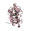
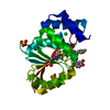
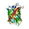
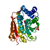
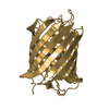
BIOMOLECULE: 1 THIS ENTRY CONTAINS THE CRYSTALLOGRAPHIC ASYMMETRIC UNIT WHICH CONSISTS OF 1 CHAIN(S) ...BIOMOLECULE: 1 THIS ENTRY CONTAINS THE CRYSTALLOGRAPHIC ASYMMETRIC UNIT WHICH CONSISTS OF 1 CHAIN(S). UNDER PHYSIOLOGICAL CONDITIONS TL5A FORMS HEXAMERIC OR OCTAMERIC ARRANGEMENTS, BUT THE PACKING DOES NOT ALLOW CREATION OF HEXAMERIC OR OCTAMERIC OLIGOMERIZATION.
Resolution: 2.01→2.13 Å / Redundancy: 5.2 % / Rmerge(I) obs: 0.344 / % possible all: 94.6
-
Processing
Software
Name
Version
Classification
MOSFLM
datareduction
SCALA
datascaling
CCP4
modelbuilding
CNS
1
refinement
CCP4
(SCALA)
datascaling
CCP4
phasing
Refinement
Method to determine structure: MIR / Resolution: 2.01→15.25 Å / Isotropic thermal model: RESTRAINED / Cross valid method: THROUGHOUT / σ(F): 0 / σ(I): 0 / Stereochemistry target values: ENGH & HUBER Details: 44 residues N-terminal and 5 residues C-terminal could not be resolved due to badly defined electron density.
Rfactor
Num. reflection
% reflection
Selection details
Rfree
0.198
2412
-
RANDOM
Rwork
0.183
-
-
-
all
-
48257
-
-
obs
-
48257
98.1 %
-
Displacement parameters
Biso mean: 27.95 Å2
Baniso -1
Baniso -2
Baniso -3
1-
1.35 Å2
0 Å2
0 Å2
2-
-
1.35 Å2
0 Å2
3-
-
-
-2.7 Å2
Refine analyze
Free
Obs
Luzzati coordinate error
0.24 Å
0.22 Å
Luzzati d res low
-
5 Å
Luzzati sigma a
0.29 Å
6.29 Å
Refinement step
Cycle: LAST / Resolution: 2.01→15.25 Å
Protein
Nucleic acid
Ligand
Solvent
Total
Num. atoms
1839
0
16
179
2034
Refine LS restraints
Refine-ID
Type
Dev ideal
X-RAY DIFFRACTION
c_bond_d
0.005
X-RAY DIFFRACTION
c_angle_deg
1.4
X-RAY DIFFRACTION
c_dihedral_angle_d
24.3
X-RAY DIFFRACTION
c_improper_angle_d
0.7
X-RAY DIFFRACTION
c_mcbond_it
1.41
X-RAY DIFFRACTION
c_mcangle_it
2.01
LS refinement shell
Resolution: 2.01→2.13 Å / Rfactor Rfree error: 0.017
In the structure databanks used in Yorodumi, some data are registered as the other names, "COVID-19 virus" and "2019-nCoV". Here are the details of the virus and the list of structure data.
Jan 31, 2019. EMDB accession codes are about to change! (news from PDBe EMDB page)
EMDB accession codes are about to change! (news from PDBe EMDB page)
The allocation of 4 digits for EMDB accession codes will soon come to an end. Whilst these codes will remain in use, new EMDB accession codes will include an additional digit and will expand incrementally as the available range of codes is exhausted. The current 4-digit format prefixed with “EMD-” (i.e. EMD-XXXX) will advance to a 5-digit format (i.e. EMD-XXXXX), and so on. It is currently estimated that the 4-digit codes will be depleted around Spring 2019, at which point the 5-digit format will come into force.
The EM Navigator/Yorodumi systems omit the EMD- prefix.
Related info.:Q: What is EMD? / ID/Accession-code notation in Yorodumi/EM Navigator
Yorodumi is a browser for structure data from EMDB, PDB, SASBDB, etc.
This page is also the successor to EM Navigator detail page, and also detail information page/front-end page for Omokage search.
The word "yorodu" (or yorozu) is an old Japanese word meaning "ten thousand". "mi" (miru) is to see.
Related info.:EMDB / PDB / SASBDB / Comparison of 3 databanks / Yorodumi Search / Aug 31, 2016. New EM Navigator & Yorodumi / Yorodumi Papers / Jmol/JSmol / Function and homology information / Changes in new EM Navigator and Yorodumi
 Movie
Movie Controller

 Yorodumi
 Open data
 Basic information
 Components
 Keywords
 Function and homology information
 Tachypleus tridentatus (Chinese horseshoe crab)
 X-RAY DIFFRACTION /
  Authors
 Citation
 Structure visualization
 Downloads & links
 Other downloads


 PDBj
PDBj


 Assembly

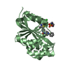
 Type: D-saccharide, beta linking / Mass: 221.208 Da / Num. of mol.: 1
 Mass: 40.078 Da / Num. of mol.: 1 / Source method: obtained synthetically / Formula: Ca
 Mass: 18.015 Da / Num. of mol.: 179 / Source method: isolated from a natural source / Formula: H2O
 Sample preparation
 Processing