 Movie
Movie Controller


+ Open data

- Basic information
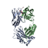
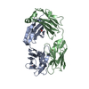
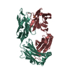
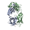
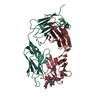
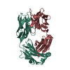
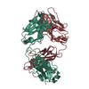
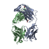
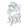
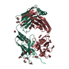
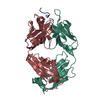
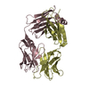
| Entry | Database: PDB / ID: 1i7z | ||||||
|---|---|---|---|---|---|---|---|
| Title | ANTIBODY GNC92H2 BOUND TO LIGAND | ||||||
 Components Components |
| ||||||
 Keywords Keywords | IMMUNE SYSTEM / IgG fold / antibody / chimera | ||||||
| Function / homology |  Function and homology information Function and homology informationIgD immunoglobulin complex / IgA immunoglobulin complex / IgM immunoglobulin complex / IgE immunoglobulin complex / Fc-gamma receptor I complex binding / CD22 mediated BCR regulation / complement-dependent cytotoxicity / IgG immunoglobulin complex / Fc epsilon receptor (FCERI) signaling / antibody-dependent cellular cytotoxicity ...IgD immunoglobulin complex / IgA immunoglobulin complex / IgM immunoglobulin complex / IgE immunoglobulin complex / Fc-gamma receptor I complex binding / CD22 mediated BCR regulation / complement-dependent cytotoxicity / IgG immunoglobulin complex / Fc epsilon receptor (FCERI) signaling / antibody-dependent cellular cytotoxicity / immunoglobulin receptor binding / immunoglobulin complex, circulating / Classical antibody-mediated complement activation / Initial triggering of complement / immunoglobulin mediated immune response / FCGR activation / Role of LAT2/NTAL/LAB on calcium mobilization / complement activation, classical pathway / Role of phospholipids in phagocytosis / Scavenging of heme from plasma / antigen binding / FCERI mediated Ca+2 mobilization / FCGR3A-mediated IL10 synthesis / Antigen activates B Cell Receptor (BCR) leading to generation of second messengers / Regulation of Complement cascade / Cell surface interactions at the vascular wall / B cell receptor signaling pathway / FCGR3A-mediated phagocytosis / FCERI mediated MAPK activation / Regulation of actin dynamics for phagocytic cup formation / FCERI mediated NF-kB activation / Immunoregulatory interactions between a Lymphoid and a non-Lymphoid cell / antibacterial humoral response / Interleukin-4 and Interleukin-13 signaling / blood microparticle / adaptive immune response / Potential therapeutics for SARS / immune response / extracellular space / extracellular exosome / extracellular region / plasma membrane Similarity search - Function | ||||||
| Biological species |   Homo sapiens (human) Homo sapiens (human) | ||||||
| Method |  X-RAY DIFFRACTION / SYNCHROTRON / MOLECULAR REPLACEMENT / Resolution: 2.3 Å X-RAY DIFFRACTION / SYNCHROTRON / MOLECULAR REPLACEMENT / Resolution: 2.3 Å | ||||||
 Authors Authors | Larsen, N.A. / Wilson, I.A. | ||||||
 Citation Citation | Journal: J.Mol.Biol. / Year: 2001 Title: Crystal structure of a cocaine-binding antibody. Authors: Larsen, N.A. / Zhou, B. / Heine, A. / Wirsching, P. / Janda, K.D. / Wilson, I.A. | ||||||
| History |
|
- Structure visualization
| Structure viewer | Molecule: MolmilJmol/JSmol |
|---|
- Downloads & links
-Download
| PDBx/mmCIF format | 1i7z.cif.gz | 180.8 KB | Display | PDBx/mmCIF format |
|---|---|---|---|---|
| PDB format | pdb1i7z.ent.gz | 144 KB | Display | PDB format |
| PDBx/mmJSON format | 1i7z.json.gz | Tree view | PDBx/mmJSON format | |
| Others |  Other downloads Other downloads |
-Validation report
| Summary document | 1i7z_validation.pdf.gz | 1 MB | Display | wwPDB validaton report |
|---|---|---|---|---|
| Full document | 1i7z_full_validation.pdf.gz | 1.1 MB | Display | |
| Data in XML | 1i7z_validation.xml.gz | 36.2 KB | Display | |
| Data in CIF | 1i7z_validation.cif.gz | 50.4 KB | Display | |
| Arichive directory | https://data.pdbj.org/pub/pdb/validation_reports/i7/1i7zftp://data.pdbj.org/pub/pdb/validation_reports/i7/1i7z | HTTPS FTP |
-Related structure data
| Related structure data |  1nsnS S: Starting model for refinement |
|---|---|
| Similar structure data |


-Links
 PDBj
PDBj


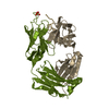
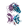
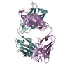
- Assembly
| Deposited unit | 
| ||||||||
|---|---|---|---|---|---|---|---|---|---|
| 1 | 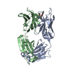
| ||||||||
| 2 | 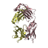
| ||||||||
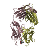
| Unit cell |
|
-Components
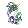
| #1: Antibody | Mass: 23951.682 Da / Num. of mol.: 2 Source method: isolated from a genetically manipulated source Details: THE CHIMERA CONSISTS OF RESIDUES 1-108 OF MOUSE PORTION AND 109-214 OF HUMAN PORTION OF IG KAPPA CHAIN. Source: (gene. exp.) Mus musculus, Homo sapiens / Genus: Mus, Homo / Species: , / Strain: , / Plasmid: PET / Species (production host): Escherichia coli / Production host:  #2: Antibody | Mass: 23571.344 Da / Num. of mol.: 2 Source method: isolated from a genetically manipulated source Details: THE CHIMERA CONSISTS OF RESIDUES 1-113 OF MOUSE PORTION AND 114-228 OF HUMAN PORTION OF IG GAMMA-1 CHAIN. Source: (gene. exp.) Mus musculus, Homo sapiens / Genus: Mus, Homo / Species: , / Strain: , / Plasmid: PET / Species (production host): Escherichia coli / Production host: #3: Chemical |   Mass: 303.353 Da / Num. of mol.: 2 / Source method: obtained synthetically / Formula: C17H21NO4 Mass: 303.353 Da / Num. of mol.: 2 / Source method: obtained synthetically / Formula: C17H21NO4#4: Water | ChemComp-HOH / |  Mass: 18.015 Da / Num. of mol.: 233 / Source method: isolated from a natural source / Formula: H2O Mass: 18.015 Da / Num. of mol.: 233 / Source method: isolated from a natural source / Formula: H2OHas protein modification | Y | |
|---|
-Experimental details
-Experiment
| Experiment | Method: X-RAY DIFFRACTION / Number of used crystals: 1 |
|---|
- Sample preparation
| Crystal | Density Matthews: 2.59 Å3/Da / Density % sol: 52.57 % | ||||||||||||||||||||||||||||
|---|---|---|---|---|---|---|---|---|---|---|---|---|---|---|---|---|---|---|---|---|---|---|---|---|---|---|---|---|---|
| Crystal grow | *PLUS pH: 4.5 / Method: unknown | ||||||||||||||||||||||||||||
| Components of the solutions | *PLUS
|
-Data collection
| Diffraction | Mean temperature: 100 K |
|---|---|
| Diffraction source | Source: SYNCHROTRON / Site: SSRL  / Beamline: BL9-2 / Wavelength: 1.039 Å / Beamline: BL9-2 / Wavelength: 1.039 Å |
| Detector | Type: ADSC QUANTUM 4 / Detector: CCD / Date: May 20, 2000 |
| Radiation | Protocol: SINGLE WAVELENGTH / Monochromatic (M) / Laue (L): M / Scattering type: x-ray |
| Radiation wavelength | Wavelength: 1.039 Å / Relative weight: 1 |
| Reflection | Resolution: 2.3→20 Å / Num. all: 164865 / Num. obs: 43057 / % possible obs: 98.9 % / Observed criterion σ(F): 10.6 / Observed criterion σ(I): 113.1 / Redundancy: 3.8 % / Biso Wilson estimate: 30 Å2 / Rmerge(I) obs: 0.082 / Net I/σ(I): 16.9 |
| Reflection shell | Resolution: 2.3→2.34 Å / Redundancy: 3.5 % / Rmerge(I) obs: 0.512 / Mean I/σ(I) obs: 2.4 / % possible all: 85.7 |
| Reflection | *PLUS Num. measured all: 164865 |
| Reflection shell | *PLUS Highest resolution: 2.3 Å / % possible obs: 85.7 % |
- Processing
| Software |
| ||||||||||||||||||||||||
|---|---|---|---|---|---|---|---|---|---|---|---|---|---|---|---|---|---|---|---|---|---|---|---|---|---|
| Refinement | Method to determine structure: MOLECULAR REPLACEMENT Starting model: PDB ENTRY 1NSN Resolution: 2.3→20 Å / Cross valid method: THROUGHOUT / σ(F): 0 / σ(I): 0 / Stereochemistry target values: Engh & Huber
| ||||||||||||||||||||||||
| Refinement step | Cycle: LAST / Resolution: 2.3→20 Å
| ||||||||||||||||||||||||
| Refinement | *PLUS Rfactor obs: 0.219 / Rfactor Rwork: 0.219 | ||||||||||||||||||||||||
| Solvent computation | *PLUS | ||||||||||||||||||||||||
| Displacement parameters | *PLUS | ||||||||||||||||||||||||
| Refine LS restraints | *PLUS
|
