 Movie
Movie Controller

[English] 日本語
 Yorodumi
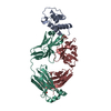
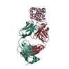
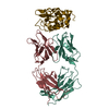
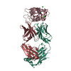
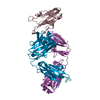
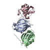
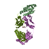
- PDB-1nsn: THE CRYSTAL STRUCTURE OF ANTIBODY N10-STAPHYLOCOCCAL NUCLEASE COM... -
+ Open data

- Basic information
| Entry | Database: PDB / ID: 1nsn | ||||||
|---|---|---|---|---|---|---|---|
| Title | THE CRYSTAL STRUCTURE OF ANTIBODY N10-STAPHYLOCOCCAL NUCLEASE COMPLEX AT 2.9 ANGSTROMS RESOLUTION | ||||||
 Components Components |
| ||||||
 Keywords Keywords | COMPLEX (IMMUNOGLOBULIN/HYDROLASE) / IMMUNOGLOBULIN / STAPHYLOCOCCAL NUCLEASE / COMPLEX (IMMUNOGLOBULIN-HYDROLASE) COMPLEX | ||||||
| Function / homology |  Function and homology information Function and homology informationmicrococcal nuclease / 3' overhang single-stranded DNA endonuclease activity / nucleic acid binding / extracellular region / membrane / metal ion binding Similarity search - Function | ||||||
| Biological species |   Staphylococcus aureus (bacteria) Staphylococcus aureus (bacteria) | ||||||
| Method |  X-RAY DIFFRACTION / Resolution: 2.8 Å X-RAY DIFFRACTION / Resolution: 2.8 Å | ||||||
 Authors Authors | Sheriff, S. / Bossart-Whitaker, P. | ||||||
 Citation Citation | Journal: J.Mol.Biol. / Year: 1995 Title: The crystal structure of the antibody N10-staphylococcal nuclease complex at 2.9 A resolution. Authors: Bossart-Whitaker, P. / Chang, C.Y. / Novotny, J. / Benjamin, D.C. / Sheriff, S. #1: Journal: J.Mol.Biol. / Year: 1994Title: Crystallization and Preliminary X-Ray Analysis of an Anti-Staphylococcal Nuclease-Staphylococcal Nuclease Complex and of a Second Anti-Staphylococcal Nuclease Antibody Authors: Chang, C.Y. / Bossart-Whitaker, P. / Tabernero, L. / Einspahr, H. / Workman, L. / Benjamin, D.C. / Sheriff, S. | ||||||
| History |
|
- Structure visualization
| Structure viewer | Molecule: MolmilJmol/JSmol |
|---|
- Downloads & links
-Download
| PDBx/mmCIF format | 1nsn.cif.gz | 120.4 KB | Display | PDBx/mmCIF format |
|---|---|---|---|---|
| PDB format | pdb1nsn.ent.gz | 93.1 KB | Display | PDB format |
| PDBx/mmJSON format | 1nsn.json.gz | Tree view | PDBx/mmJSON format | |
| Others |  Other downloads Other downloads |
-Validation report
| Arichive directory | https://data.pdbj.org/pub/pdb/validation_reports/ns/1nsnftp://data.pdbj.org/pub/pdb/validation_reports/ns/1nsn | HTTPS FTP |
|---|
-Related structure data
| Similar structure data |
|---|


-Links
 PDBj
PDBj

- Assembly
| Deposited unit | 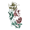
| ||||||||
|---|---|---|---|---|---|---|---|---|---|
| 1 |
| ||||||||
| Unit cell |
| ||||||||
| Atom site foot note | 1: CIS PROLINE - PRO L 8 / 2: CIS PROLINE - PRO L 95 / 3: CIS PROLINE - PRO H 149 / 4: CIS PROLINE - PRO H 151 / 5: CIS PROLINE - PRO H 202 / 6: CIS PROLINE - PRO S 117 |
-Components
| #1: Antibody | Mass: 24189.666 Da / Num. of mol.: 1 / Source method: isolated from a natural source / Source: (natural) |
|---|---|
| #2: Antibody | Mass: 22580.119 Da / Num. of mol.: 1 / Source method: isolated from a natural source / Source: (natural) |
| #3: Protein | Mass: 16843.330 Da / Num. of mol.: 1 Source method: isolated from a genetically manipulated source Source: (gene. exp.) Staphylococcus aureus (bacteria) / Strain: FOGGI / Gene: NUCLEASE / Plasmid: PFOG405 / Gene (production host): NUCLEASE / Production host: |
| Has protein modification | Y |
-Experimental details
-Experiment
| Experiment | Method: X-RAY DIFFRACTION / Number of used crystals: 1 |
|---|
- Sample preparation
| Crystal | Density Matthews: 2.86 Å3/Da / Density % sol: 57.03 % | ||||||||||||||||||||
|---|---|---|---|---|---|---|---|---|---|---|---|---|---|---|---|---|---|---|---|---|---|
| Crystal grow | *PLUS Method: vapor diffusion, hanging drop / Details: Chang, C.Y., (1994) J.Mol.Biol., 239, 154. | ||||||||||||||||||||
| Components of the solutions | *PLUS
|
-Data collection
| Diffraction source | Wavelength: 1.5418 Å |
|---|---|
| Detector | Type: SIEMENS / Detector: AREA DETECTOR / Date: Nov 1, 1992 |
| Radiation | Monochromatic (M) / Laue (L): M / Scattering type: x-ray |
| Radiation wavelength | Wavelength: 1.5418 Å / Relative weight: 1 |
| Reflection | Resolution: 2.7→38 Å / Num. obs: 14626 / % possible obs: 78.7 % / Observed criterion σ(I): 0 / Redundancy: 2 % / Rmerge(I) obs: 0.048 |
| Reflection | *PLUS % possible obs: 81 % / Num. measured all: 32555 / Rmerge(I) obs: 0.048 |
| Reflection shell | *PLUS Highest resolution: 2.77 Å / Lowest resolution: 2.89 Å / % possible obs: 25 % / Rmerge(I) obs: 0.103 |
- Processing
| Software |
| ||||||||||||||||||||||||||||||||||||||||||||||||||||||||||||
|---|---|---|---|---|---|---|---|---|---|---|---|---|---|---|---|---|---|---|---|---|---|---|---|---|---|---|---|---|---|---|---|---|---|---|---|---|---|---|---|---|---|---|---|---|---|---|---|---|---|---|---|---|---|---|---|---|---|---|---|---|---|
| Refinement | Resolution: 2.8→5 Å / σ(F): 0
| ||||||||||||||||||||||||||||||||||||||||||||||||||||||||||||
| Displacement parameters | Biso mean: 18 Å2 | ||||||||||||||||||||||||||||||||||||||||||||||||||||||||||||
| Refine analyze | Luzzati coordinate error obs: 0.34 Å | ||||||||||||||||||||||||||||||||||||||||||||||||||||||||||||
| Refinement step | Cycle: LAST / Resolution: 2.8→5 Å
| ||||||||||||||||||||||||||||||||||||||||||||||||||||||||||||
| Refine LS restraints |
| ||||||||||||||||||||||||||||||||||||||||||||||||||||||||||||
| Software | *PLUS Name: X-PLOR / Version: 3.1 / Classification: refinement | ||||||||||||||||||||||||||||||||||||||||||||||||||||||||||||
| Refinement | *PLUS Lowest resolution: 5 Å / Rfactor Rfree: 0.284 | ||||||||||||||||||||||||||||||||||||||||||||||||||||||||||||
| Solvent computation | *PLUS | ||||||||||||||||||||||||||||||||||||||||||||||||||||||||||||
| Displacement parameters | *PLUS Biso mean: 19 Å2 | ||||||||||||||||||||||||||||||||||||||||||||||||||||||||||||
| Refine LS restraints | *PLUS
|
