 ムービー
ムービー コントローラー
 構造ビューア
 EMN検索について


-検索条件
-検索結果
検索 (著者・登録者: chen & yh)の結果97件中、1から50件目までを表示しています

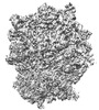
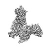
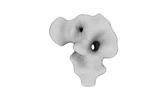
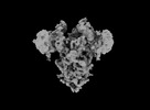
EMDB-37604: 
Structural basis of translation inhibition by a valine tRNA-derived fragment

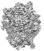
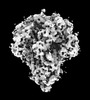
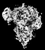
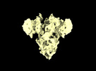
EMDB-37733:
Structural basis of translation inhibition by a valine tRNA-derived fragment

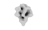
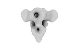
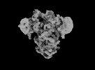
EMDB-37734:
Structural basis of translation inhibition by a valine tRNA-derived fragment
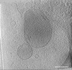
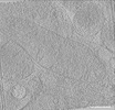
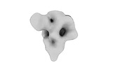
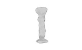
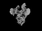
EMDB-39012:
Representative tomogram of primary glioblastoma stem cell with circular inter-mitochondrial junctions.
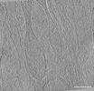
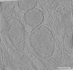
EMDB-39015:
Representative tomogram of microglia cell with nanotunnel-like structures resembling mitochondrial fission.
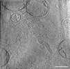
EMDB-39019:
Representative tomogram of glioblastoma cell with nanotunnel-like structure and inter-mitochondrial junction.
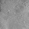
EMDB-39021:
Representative tomogram of normal human astrocyte with nanotunnel-like structure which is an extension of the mitochondrial outer membrane.
EMDB-39023:
Representative tomogram of primary glioblastoma differentiated cell with parallel inter-mitochondrial junction.
EMDB-39024:
Representative tomogram of primary glioblastoma stem cell with clustered mitochondria bearing various long-short axis ratios.
EMDB-34866:
Cryo-EM Structures and Translocation Mechanism of Crenarchaeota Ribosome
EMDB-34869:
Cryo-EM Structures and Translocation Mechanism of Crenarchaeota Ribosome

EMDB-35775:
The rice Na+/H+ antiporter SOS1 in an auto-inhibited state

EMDB-35950:
The truncated rice Na+/H+ antiporter SOS1 (1-976) in a constitutively active state
EMDB-34870:
Cryo-EM Structures and Translocation Mechanism of Crenarchaeota Ribosome
EMDB-34867:
Cryo-EM Structures and Translocation Mechanism of Crenarchaeota Ribosome
EMDB-34868:
Cryo-EM Structures and Translocation Mechanism of Crenarchaeota Ribosome
EMDB-34863:
Cryo-EM Structures and Translocation Mechanism of Crenarchaeota Ribosome
EMDB-34864:
Cryo-EM Structures and Translocation Mechanism of Crenarchaeota Ribosome
EMDB-34860:
Cryo-EM Structures and Translocation Mechanism of Crenarchaeota Ribosome

EMDB-34861:
Cryo-EM Structures and Translocation Mechanism of Crenarchaeota Ribosome

EMDB-34862:
Cryo-EM Structures and Translocation Mechanism of Crenarchaeota Ribosome

EMDB-34710:
Cryo-EM structure of WeiTsing

EMDB-33145:
Cryo-EM structures of human mitochondrial NAD(P)+-dependent malic enzyme in apo form

EMDB-33146:
Cryo-EM structures of human mitochondrial NAD(P)+-dependent malic enzyme in a ternary complex with NAD+ and allosteric inhibitor EA

EMDB-33147:
Cryo-EM structures of human mitochondrial NAD(P)+-dependent malic enzyme in a ternary complex with NAD+ and allosteric inhibitor MDSA
EMDB-35522:
Cryo-EM structure of the TUG891 bound GPR120-Giq complex(mask on receptor)
EMDB-35523:
Cryo-EM structure of the TUG891 bound GPR120-Giq complex(mask on Giq-scFV16 complex)
EMDB-35524:
Cryo-EM structure of the eicosapentaenoic acid bound GPR120-Gi1 complex(mask on receptor)
EMDB-35525:
Cryo-EM structure of the eicosapentaenoic acid bound GPR120-Gi1 complex(mask on Gil-scFV16 complex)
EMDB-35529:
Cryo-EM structure of the TUG891 bound GPR120-Giq complex (consensus map)
EMDB-35533:
Cryo-EM structure of the eicosapentaenoic acid bound GPR120-Gi complex(consensus map)

EMDB-35356:
Cryo-EM structure of the 9-hydroxystearic acid bound GPR120-Gi complex

EMDB-35357:
Cryo-EM structure of the linoleic acid bound GPR120-Gi complex

EMDB-35358:
Cryo-EM structure of the oleic acid bound GPR120-Gi complex

EMDB-35359:
Cryo-EM structure of the TUG891 bound GPR120-Gi complex

EMDB-35360:
Cryo-EM structure of the eicosapentaenoic acid bound GPR120-Gi complex
EMDB-29736:
Cryo-EM structure of the TUG891 bound GPR120-Giq complex

EMDB-33923:
Cryo-EM structure of SARS-CoV-2 Omicron spike glycoprotein in complex with three neutralizing nanobody 3-2A2-4
EMDB-32329:
Cryo-EM map of PEDV (Pintung 52) S protein with all three protomers in the D0-down conformation determined in situ on intact viral particles.
EMDB-32332:
Subtomogram averaging of PEDV (Pintung 52) S protein with all three protomers in the D0-down conformation determined in situ on intact viral particles.
EMDB-32333:
Subtomogram averaging of PEDV (Pintung 52) S protein with one protomer in the D0-up conformation and two protomers in the D0-down conformation, determined in situ on intact viral particles
EMDB-32337:
Subtomogram averaging of PEDV (Pintung 52) S protein with two protomers in the D0-up conformation and one protomer in the D0-down conformation, determined in situ on intact viral particles.
EMDB-32338:
Cryo-EM map of PEDV S protein with one protomer in the D0-up conformation while the other two in the D0-down conformation
EMDB-32339:
Subtomogram averaging of PEDV (Pintung 52) S protein with all three protomers in the D0-up conformation determined in situ on intact viral particles.
EMDB-32340:
Subtomogram averaging of PEDV (Pintung 52) S protein in the postfusion form determined in situ on intact viral particles.
EMDB-33646:
Cryo-EM map of IPEC-J2 cell-derived PEDV PT52 S protein with three D0-up
EMDB-33647:
Cryo-EM map of IPEC-J2 cell-derived PEDV PT52 S protein one D0-down and two D0-up
EMDB-33648:
Symmetry-expanded and locally refined protomer structure of IPEC-J2 cell-derived PEDV PT52 S with a CTD-close conformation
EMDB-33649:
Symmetry-expanded and locally refined protomer structure of IPEC-J2 cell-derived PEDV PT52 S with a CTD-open conformation
EMDB-33700:
Cryo-EM map of HEK293F cell-derived PEDV PT52 S protein with three D0-down
ページ:
 wwPDBはEMDBデータモデルのバージョン3へ移行します
