 Movie
Movie Controller
 Structure viewers
 About EMN search


-Search query
-Search result
Showing 1 - 50 of 128 items for (author: sauer & rt)
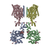
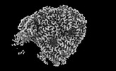
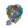
EMDB-17377: 
Structure of human SIT1 (focussed map / refinement)

EMDB-17378:
Structure of human SIT1:ACE2 complex (open PD conformation)

EMDB-17379:
Structure of human SIT1:ACE2 complex (closed PD conformation)
EMDB-17380:
Structure of human SIT1 bound to L-pipecolate (focussed map / refinement)
EMDB-17381:
Structure of human SIT1:ACE2 complex (open PD conformation) bound to L-pipecolate
EMDB-17382:
Structure of human SIT1:ACE2 complex (closed PD conformation) bound to L-pipecolate

PDB-8p2w:
Structure of human SIT1 (focussed map / refinement)

PDB-8p2x:
Structure of human SIT1:ACE2 complex (open PD conformation)

PDB-8p2y:
Structure of human SIT1:ACE2 complex (closed PD conformation)

PDB-8p2z:
Structure of human SIT1 bound to L-pipecolate (focussed map / refinement)
PDB-8p30:
Structure of human SIT1:ACE2 complex (open PD conformation) bound to L-pipecolate

PDB-8p31:
Structure of human SIT1:ACE2 complex (closed PD conformation) bound to L-pipecolate

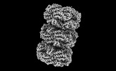
EMDB-19109:
Structure of XPD stalled at a Y-fork DNA containing a interstrand crosslink

PDB-8rev:
Structure of XPD stalled at a Y-fork DNA containing a interstrand crosslink
EMDB-41434:
Bottom cylinder of high-resolution phycobilisome quenched by OCP (local refinement)
EMDB-41435:
Central rod disk in C1 symmetry of high-resolution phycobilisome quenched by OCP (local refinement)

EMDB-41436:
Central rod disk in D3 symmetry of high-resolution phycobilisome quenched by OCP (local refinement)
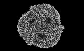
EMDB-41463:
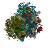
Synechocystis PCC 6803 Phycobilisome quenched by OCP, high resolution
EMDB-41475:
Top cylinder bound to OCP from high-resolution phycobilisome quenched by OCP (local refinement)
EMDB-41585:
Rod from high-resolution phycobilisome quenched by OCP (local refinement)
PDB-8to2:
Bottom cylinder of high-resolution phycobilisome quenched by OCP (local refinement)
PDB-8to5:
Central rod disk in C1 symmetry of high-resolution phycobilisome quenched by OCP (local refinement)
PDB-8tpj:
Top cylinder bound to OCP from high-resolution phycobilisome quenched by OCP (local refinement)

PDB-8tro:
Rod from high-resolution phycobilisome quenched by OCP (local refinement)

EMDB-18659:
Cryo-EM Structure of Human Kv3.1 in Complex with Modulator AUT1

EMDB-18660:
Cryo-EM Structure of Human Kv3.1 in Complex with Modulator AUT5

PDB-8quc:
Cryo-EM Structure of Human Kv3.1 in Complex with Modulator AUT1

PDB-8qud:
Cryo-EM Structure of Human Kv3.1 in Complex with Modulator AUT5

EMDB-18387:
FtsH1 protease from P.aeruginosa clone C in negative stain

EMDB-18388:
FtsH2 protease from P.aeruginosa clone C in negative stain

EMDB-18389:
P.aeruginosa clone C construct PaFtsH2-H1-link32 in negative stain
EMDB-27941:
Cryo-EM structure of substrate-free DNClpX.ClpP from singly capped particles
EMDB-27946:
Cryo-EM structure of substrate-free DNClpX.ClpP
EMDB-27952:
Cryo-EM structure of substrate-free ClpX.ClpP
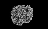
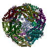
EMDB-33329:
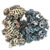
High resolution cry-EM structure of the human 80S ribosome from SNORD127+/+ Kasumi-1 cells
EMDB-33330:
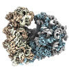
High resolution cry-EM structure of the human 80S ribosome from SNORD127+/- Kasumi-1 cells
PDB-7xnx:
High resolution cry-EM structure of the human 80S ribosome from SNORD127+/+ Kasumi-1 cells
PDB-7xny:
High resolution cry-EM structure of the human 80S ribosome from SNORD127+/- Kasumi-1 cells
EMDB-28585:
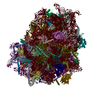
Cryo-EM structure of a delivery complex containing the SspB adaptor, an ssrA-tagged substrate, and the AAA+ ClpXP protease
PDB-8et3:
Cryo-EM structure of a delivery complex containing the SspB adaptor, an ssrA-tagged substrate, and the AAA+ ClpXP protease

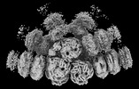
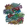
EMDB-26554:
ClpAP complex bound to ClpS N-terminal extension, class IIa

EMDB-26555:
ClpAP complex bound to ClpS N-terminal extension, class IIb

EMDB-26556:
ClpAP complex bound to ClpS N-terminal extension, class I

EMDB-26558:
ClpAP complex bound to ClpS N-terminal extension, class IIc

EMDB-26559:
ClpAP complex bound to ClpS N-terminal extension, class IIIb

PDB-7uiv:
ClpAP complex bound to ClpS N-terminal extension, class IIa

PDB-7uiw:
ClpAP complex bound to ClpS N-terminal extension, class IIb

PDB-7uix:
ClpAP complex bound to ClpS N-terminal extension, class I

PDB-7uiz:
ClpAP complex bound to ClpS N-terminal extension, class IIc

PDB-7uj0:
ClpAP complex bound to ClpS N-terminal extension, class IIIb
Pages:
 wwPDB to switch to version 3 of the EMDB data model
