Movie
Movie Controller

[English] 日本語
 Yorodumi
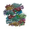
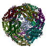
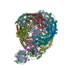
- EMDB-41436: Central rod disk in D3 symmetry of high-resolution phycobilisome ... -
+ Open data

- Basic information
| Entry |  | |||||||||||||||
|---|---|---|---|---|---|---|---|---|---|---|---|---|---|---|---|---|
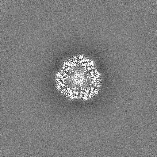
| Title | Central rod disk in D3 symmetry of high-resolution phycobilisome quenched by OCP (local refinement) | |||||||||||||||
 Map data Map data | sharpened map | |||||||||||||||
 Sample Sample |
| |||||||||||||||
 Keywords Keywords | Complex / light harvesting / pigment / PHOTOSYNTHESIS | |||||||||||||||
| Biological species |  | |||||||||||||||
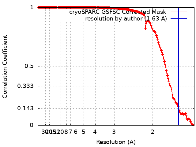
| Method | single particle reconstruction / cryo EM / Resolution: 1.63 Å | |||||||||||||||
 Authors Authors | Sauer PV / Sutter M / Cupellini L / Kirst H / Kerfeld CA | |||||||||||||||
| Funding support | European Union,  United States, United States,  Czech Republic, 4 items Czech Republic, 4 items
| |||||||||||||||
 Citation Citation | Journal: Sci Adv / Year: 2024 Title: Structural and quantum chemical basis for OCP-mediated quenching of phycobilisomes. Authors: Paul V Sauer / Lorenzo Cupellini / Markus Sutter / Mattia Bondanza / María Agustina Domínguez Martin / Henning Kirst / David Bína / Adrian Fujiet Koh / Abhay Kotecha / Basil J Greber / ...Authors: Paul V Sauer / Lorenzo Cupellini / Markus Sutter / Mattia Bondanza / María Agustina Domínguez Martin / Henning Kirst / David Bína / Adrian Fujiet Koh / Abhay Kotecha / Basil J Greber / Eva Nogales / Tomáš Polívka / Benedetta Mennucci / Cheryl A Kerfeld /    Abstract: Cyanobacteria use large antenna complexes called phycobilisomes (PBSs) for light harvesting. However, intense light triggers non-photochemical quenching, where the orange carotenoid protein (OCP) ...Cyanobacteria use large antenna complexes called phycobilisomes (PBSs) for light harvesting. However, intense light triggers non-photochemical quenching, where the orange carotenoid protein (OCP) binds to PBS, dissipating excess energy as heat. The mechanism of efficiently transferring energy from phycocyanobilins in PBS to canthaxanthin in OCP remains insufficiently understood. Using cryo-electron microscopy, we unveiled the OCP-PBS complex structure at 1.6- to 2.1-angstrom resolution, showcasing its inherent flexibility. Using multiscale quantum chemistry, we disclosed the quenching mechanism. Identifying key protein residues, we clarified how canthaxanthin's transition dipole moment in its lowest-energy dark state becomes large enough for efficient energy transfer from phycocyanobilins. Our energy transfer model offers a detailed understanding of the atomic determinants of light harvesting regulation and antenna architecture in cyanobacteria. | |||||||||||||||
| History |
|
- Structure visualization
| Supplemental images |
|---|

- Downloads & links
-EMDB archive
| Map data | emd_41436.map.gz | 483.5 MB |  EMDB map data format EMDB map data format | |
|---|---|---|---|---|
| Header (meta data) | emd-41436-v30.xmlemd-41436.xml | 18.4 KB 18.4 KB | Display Display | EMDB header |
| FSC (resolution estimation) | emd_41436_fsc.xml | 16.7 KB | Display | FSC data file |
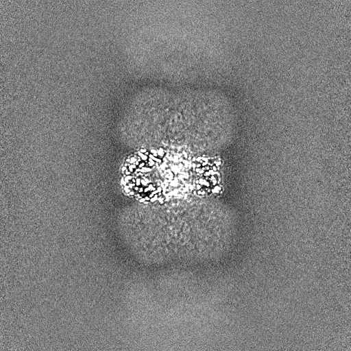
| Images | 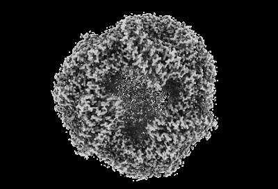 emd_41436.png emd_41436.png | 84.7 KB | ||
| Masks | emd_41436_msk_1.map | 512 MB | Mask map | |
| Filedesc metadata | emd-41436.cif.gz | 5.1 KB | ||
| Others | emd_41436_additional_1.map.gzemd_41436_half_map_1.map.gzemd_41436_half_map_2.map.gz | 258.3 MB 475.4 MB 475.4 MB | ||
| Archive directory |  http://ftp.pdbj.org/pub/emdb/structures/EMD-41436ftp://ftp.pdbj.org/pub/emdb/structures/EMD-41436 http://ftp.pdbj.org/pub/emdb/structures/EMD-41436ftp://ftp.pdbj.org/pub/emdb/structures/EMD-41436 | HTTPS FTP |
-Related structure data


-Links
| EMDB pages | EMDB (EBI/PDBe) / EMDataResource |
|---|
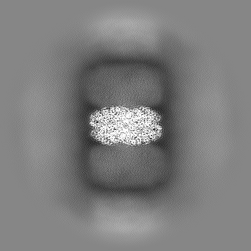
-Map
| File | Download / File: emd_41436.map.gz / Format: CCP4 / Size: 512 MB / Type: IMAGE STORED AS FLOATING POINT NUMBER (4 BYTES) | ||||||||||||||||||||||||||||||||||||
|---|---|---|---|---|---|---|---|---|---|---|---|---|---|---|---|---|---|---|---|---|---|---|---|---|---|---|---|---|---|---|---|---|---|---|---|---|---|
| Annotation | sharpened map | ||||||||||||||||||||||||||||||||||||
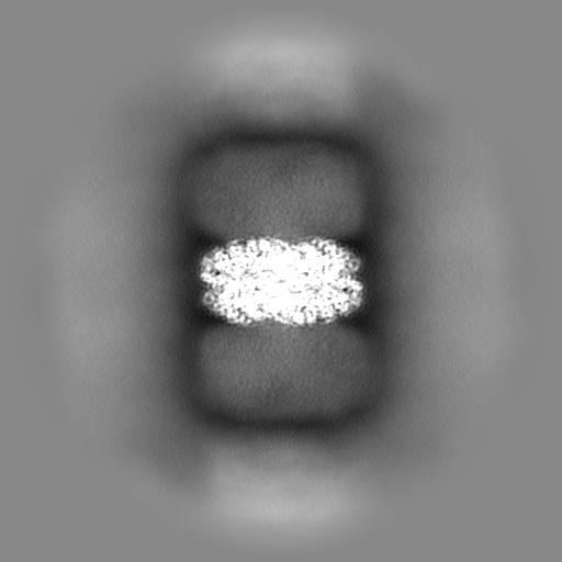
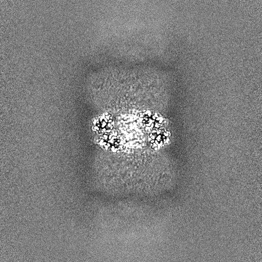
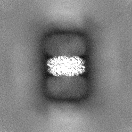
| Projections & slices | Image control
Images are generated by Spider. | ||||||||||||||||||||||||||||||||||||
| Voxel size | X=Y=Z: 0.727 Å | ||||||||||||||||||||||||||||||||||||
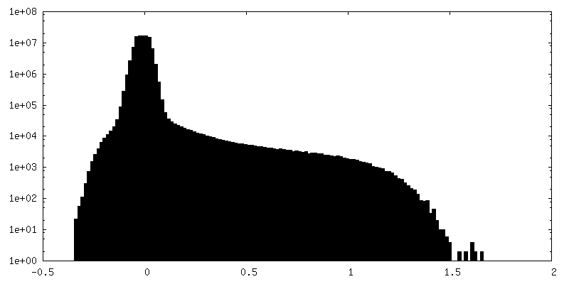
| Density |
| ||||||||||||||||||||||||||||||||||||
| Symmetry | Space group: 1 | ||||||||||||||||||||||||||||||||||||
| Details | EMDB XML:
|
 Z (Sec.)
 Y (Row.)
 X (Col.)


-Supplemental data
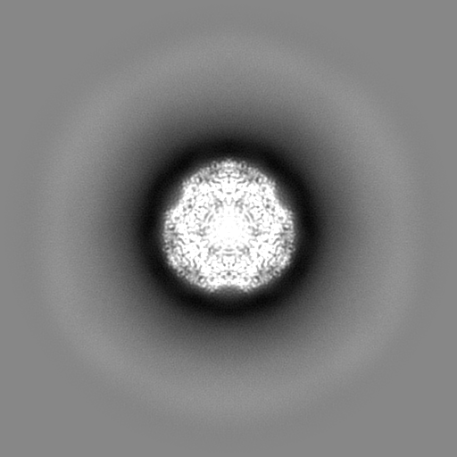
-Mask #1
| File | emd_41436_msk_1.map | ||||||||||||
|---|---|---|---|---|---|---|---|---|---|---|---|---|---|
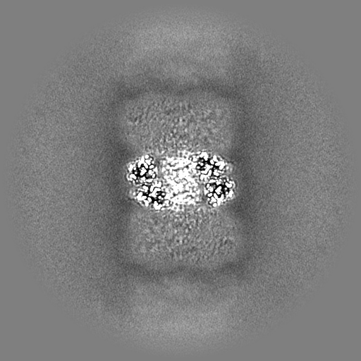
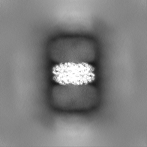
| Projections & Slices |
| ||||||||||||
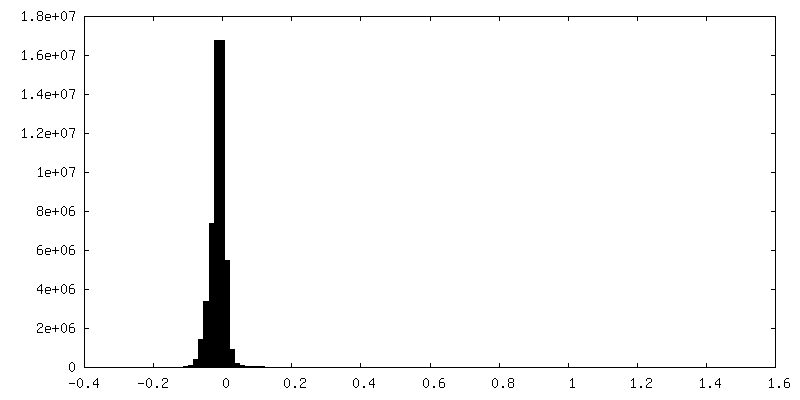
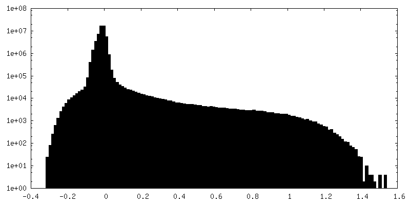
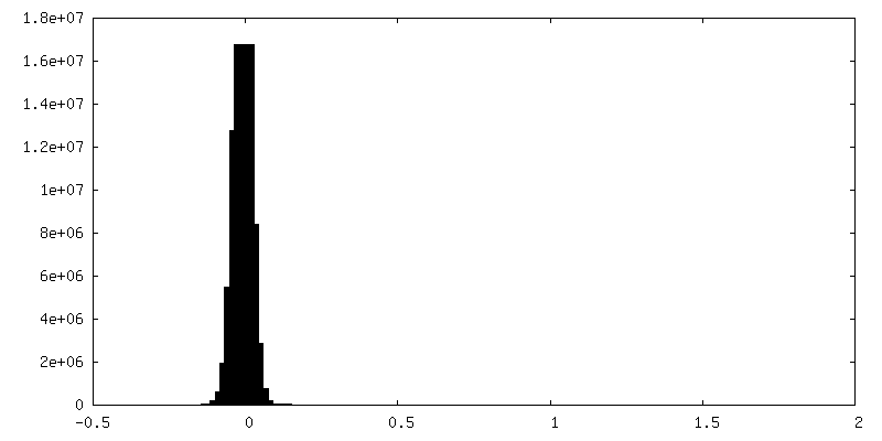
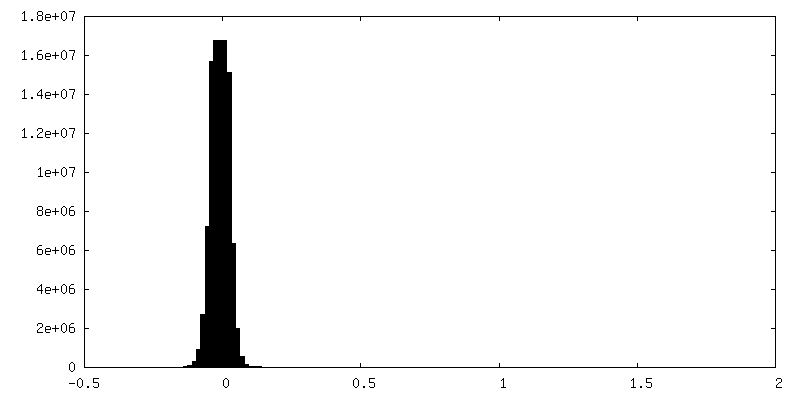
| Density Histograms |


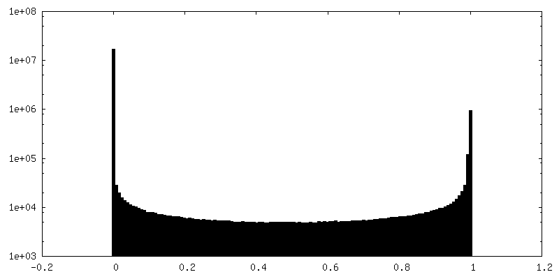
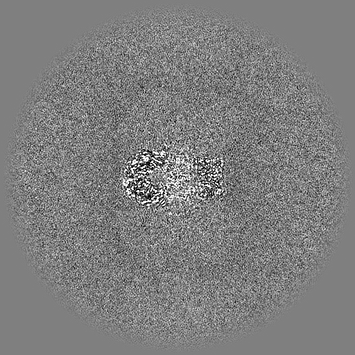
-Additional map: refined map
| File | emd_41436_additional_1.map | ||||||||||||
|---|---|---|---|---|---|---|---|---|---|---|---|---|---|
| Annotation | refined map | ||||||||||||
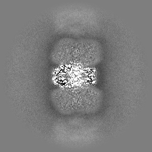
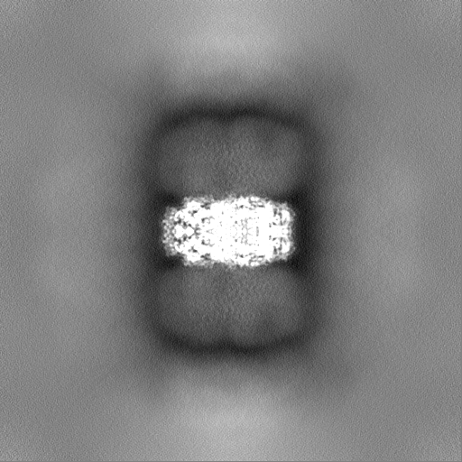
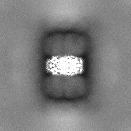
| Projections & Slices |
| ||||||||||||
| Density Histograms |


-Half map: #1
| File | emd_41436_half_map_1.map | ||||||||||||
|---|---|---|---|---|---|---|---|---|---|---|---|---|---|
| Projections & Slices |
| ||||||||||||
| Density Histograms |


-Half map: #2
| File | emd_41436_half_map_2.map | ||||||||||||
|---|---|---|---|---|---|---|---|---|---|---|---|---|---|
| Projections & Slices |
| ||||||||||||
| Density Histograms |


- Sample components
-Entire : Complex of phycobilisome from Synechocystis PCC 6803 bound to OCP
| Entire | Name: Complex of phycobilisome from Synechocystis PCC 6803 bound to OCP |
|---|---|
| Components |
|
-Supramolecule #1: Complex of phycobilisome from Synechocystis PCC 6803 bound to OCP
| Supramolecule | Name: Complex of phycobilisome from Synechocystis PCC 6803 bound to OCP type: complex / ID: 1 / Parent: 0 / Macromolecule list: all |
|---|---|
| Source (natural) | Organism: |
-Macromolecule #1: C-phycocyanin alpha subunit
| Macromolecule | Name: C-phycocyanin alpha subunit / type: protein_or_peptide / ID: 1 / Enantiomer: LEVO |
|---|---|
| Source (natural) | Organism: |
| Sequence | String: MKTPLTEAVS TADSQGRFLS STELQIAFGR LRQANAGLQA AKALTDNAQS LVNGAAQAVY NKFPYTTQTQ GNNFAADQRG KDKCARDIGY YLRIVTYCLV AGGTGPLDEY LIAGIDEINR TFDLSPSWYV EALKYIKANH GLSGDARDEA NSYLDYAINA LS |
-Macromolecule #2: C-phycocyanin beta subunit
| Macromolecule | Name: C-phycocyanin beta subunit / type: protein_or_peptide / ID: 2 / Enantiomer: LEVO |
|---|---|
| Source (natural) | Organism: |
| Sequence | String: MFDVFTRVVS QADARGEYLS GSQLDALSAT VAEGNKRIDS VNRITGNASA IVSNAARALF AEQPQLIQPG GNAYTSRRMA ACLRDMEIIL RYVTYATFTG DASVLEDRCL NGLRETYVAL GVPGASVAAG VQKMKEAALD IVNDPNGITR GDCSAIVAEI AGYFDRAAAA VA |
-Experimental details
-Structure determination
| Method | cryo EM |
|---|---|
 Processing Processing | single particle reconstruction |
| Aggregation state | particle |
-Sample preparation
| Concentration | 3.8 mg/mL |
|---|---|
| Buffer | pH: 7.5 |
| Vitrification | Cryogen name: ETHANE-PROPANE / Instrument: FEI VITROBOT MARK IV / Details: Manual blotting. |
- Electron microscopy
| Microscope | TFS KRIOS |
|---|---|
| Image recording | Film or detector model: FEI FALCON IV (4k x 4k) / Average electron dose: 40.0 e/Å2 |
| Electron beam | Acceleration voltage: 300 kV / Electron source:  FIELD EMISSION GUN FIELD EMISSION GUN |
| Electron optics | Illumination mode: OTHER / Imaging mode: BRIGHT FIELD / Nominal defocus max: 1.2 µm / Nominal defocus min: 0.4 µm |
| Sample stage | Cooling holder cryogen: NITROGEN |
| Experimental equipment |  Model: Titan Krios / Image courtesy: FEI Company |