 ムービー
ムービー コントローラー
 構造ビューア
 EMN検索について


-検索条件
-検索結果
検索 (著者・登録者: ni & tw)の結果203件中、1から50件目までを表示しています

EMDB-43275: 
GluA2 bound to GYKI-52466 and Glutamate, Inhibited State 1

EMDB-43276:
GluA2 bound to GYKI-52466 and Glutamate, Inhibited State 2

PDB-8vj6:
GluA2 bound to GYKI-52466 and Glutamate, Inhibited State 1

PDB-8vj7:
GluA2 bound to GYKI-52466 and Glutamate, Inhibited State 2
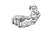
EMDB-41078:
Acinetobacter baumannii 118362 family 2A cargo-loaded encapsulin shell

EMDB-28198:
Cryo-EM map of SARS-CoV-2 Omicron BA.2 spike in complex with LLNL-199

EMDB-28199:
Cryo-EM map of SARS-CoV-2 Omicron BA.2 spike in complex with 2130-1-0114-112
EMDB-40984:
5TU-t1 - heterodimeric triplet polymerase ribozyme

PDB-8t2p:
5TU-t1 - heterodimeric triplet polymerase ribozyme
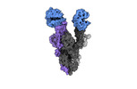
EMDB-40976:
Cryo-EM structure of mink variant Y453F trimeric spike protein bound to two mink ACE2 receptors

EMDB-40977:
Cryo-EM structure of mink variant Y453F trimeric spike protein
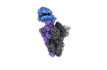
EMDB-40978:
Cryo-EM structure of mink variant Y453F trimeric spike protein bound to one mink ACE2 receptors at downRBD conformation

EMDB-40979:
Cryo-EM structure of the RBD-ACE2 interface of the SARS-CoV-2 trimeric spike protein bound to ACE2 receptor after local refinement at upRBD conformation

EMDB-40980:
Cryo-EM structure of the RBD-ACE2 interface of the SARS-CoV-2 trimeric spike protein bound to ACE2 receptor after local refinement at downRBD conformation.

EMDB-41143:
Cryo-EM structure of mink variant Y453F trimeric spike protein bound to one mink ACE2 receptors

PDB-8t20:
Cryo-EM structure of mink variant Y453F trimeric spike protein bound to two mink ACE2 receptors

PDB-8t21:
Cryo-EM structure of mink variant Y453F trimeric spike protein

PDB-8t22:
Cryo-EM structure of mink variant Y453F trimeric spike protein bound to one mink ACE2 receptors at downRBD conformation

PDB-8t23:
Cryo-EM structure of the RBD-ACE2 interface of the SARS-CoV-2 trimeric spike protein bound to ACE2 receptor after local refinement at upRBD conformation

PDB-8t25:
Cryo-EM structure of the RBD-ACE2 interface of the SARS-CoV-2 trimeric spike protein bound to ACE2 receptor after local refinement at downRBD conformation.

PDB-8taz:
Cryo-EM structure of mink variant Y453F trimeric spike protein bound to one mink ACE2 receptors

EMDB-29605:
TpeA bound closed MthK-A88F mutant in nanodisc
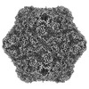
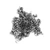
EMDB-16978:
Architecture of native chromatin fibres revealed by cryo-ET in situ
EMDB-16979:
Architecture of native chromatin fibres revealed by cryo-ET in situ
EMDB-16980:
Architecture of native chromatin fibres revealed by cryo-ET in situ

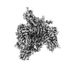
EMDB-29813:
mtHsp60 V72I apo

EMDB-29814:
mtHsp60 V72I apo focus

EMDB-29815:
ATP-bound mtHsp60 V72I

EMDB-29816:
ATP-bound mtHsp60 V72I focus

EMDB-29817:
ATP- and mtHsp10-bound mtHsp60 V72I

EMDB-29818:
ATP- and mtHsp10-bound mtHsp60 V72I focus

PDB-8g7j:
mtHsp60 V72I apo

PDB-8g7k:
mtHsp60 V72I apo focus

PDB-8g7l:
ATP-bound mtHsp60 V72I

PDB-8g7m:
ATP-bound mtHsp60 V72I focus

PDB-8g7n:
ATP- and mtHsp10-bound mtHsp60 V72I

PDB-8g7o:
ATP- and mtHsp10-bound mtHsp60 V72I focus

EMDB-27459:
MthK-A90L mutant in closed state with 0 Ca2+
EMDB-28776:
Structure of VSD4-NaV1.7-NaVPas channel chimera bound to the hybrid inhibitor GNE-1305
EMDB-28777:
Structure of VSD4-NaV1.7-NaVPas channel chimera bound to the acylsulfonamide inhibitor GDC-0310
EMDB-28778:
Structure of VSD4-NaV1.7-NaVPas channel chimera bound to the arylsulfonamide inhibitor GNE-3565

EMDB-28779:
Structure of VSD4-NaV1.7-NaVPas channel chimera bound to the hybrid inhibitor GNE-9296

PDB-8f0p:
Structure of VSD4-NaV1.7-NaVPas channel chimera bound to the hybrid inhibitor GNE-1305

PDB-8f0q:
Structure of VSD4-NaV1.7-NaVPas channel chimera bound to the acylsulfonamide inhibitor GDC-0310

PDB-8f0r:
Structure of VSD4-NaV1.7-NaVPas channel chimera bound to the arylsulfonamide inhibitor GNE-3565

PDB-8f0s:
Structure of VSD4-NaV1.7-NaVPas channel chimera bound to the hybrid inhibitor GNE-9296
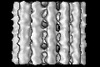
EMDB-15532:
Plasmodium falciparum sporozoite subpellicular microtubule with interrupted luminal helix determined in situ
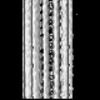
EMDB-15534:
Plasmodium falciparum gametocyte subpellicular microtubule with 13 protofilaments determined in situ
EMDB-15535:
Plasmodium falciparum gametocyte subpellicular microtubule with 14 protofilaments determined in situ

EMDB-15536:
Plasmodium falciparum gametocyte subpellicular microtubule with 15 protofilaments determined in situ
ページ:
 wwPDBはEMDBデータモデルのバージョン3へ移行します
