 Movie
Movie Controller

[English] 日本語
 Yorodumi
- EMDB-15535: Plasmodium falciparum gametocyte subpellicular microtubule with 1... -
+ Open data

- Basic information
| Entry | 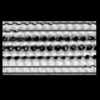 | ||||||||||||
|---|---|---|---|---|---|---|---|---|---|---|---|---|---|
| Title | Plasmodium falciparum gametocyte subpellicular microtubule with 14 protofilaments determined in situ | ||||||||||||
 Map data Map data | Plasmodium falciparum Gametocyte subpellicular microtubule with 14 protofilaments | ||||||||||||
 Sample Sample |
| ||||||||||||
 Keywords Keywords | Cytoskeleton / microtubules / STRUCTURAL PROTEIN | ||||||||||||
| Biological species |  | ||||||||||||
| Method | subtomogram averaging / cryo EM / Resolution: 24.0 Å | ||||||||||||
 Authors Authors | Ferreira JL / Prazak V / Vasishtan D / Grunewald K / Siggel M / Hentzschel F / Pietsch E / Kosinski J / Frischknecht F / Gilberger TW | ||||||||||||
| Funding support |  France, France,  United Kingdom, United Kingdom,  Germany, 3 items Germany, 3 items
| ||||||||||||
 Citation Citation | Journal: Nat Commun / Year: 2023 Title: Variable microtubule architecture in the malaria parasite. Authors: Josie L Ferreira / Vojtěch Pražák / Daven Vasishtan / Marc Siggel / Franziska Hentzschel / Annika M Binder / Emma Pietsch / Jan Kosinski / Friedrich Frischknecht / Tim W Gilberger / Kay Grünewald / Abstract: Microtubules are a ubiquitous eukaryotic cytoskeletal element typically consisting of 13 protofilaments arranged in a hollow cylinder. This arrangement is considered the canonical form and is adopted ...Microtubules are a ubiquitous eukaryotic cytoskeletal element typically consisting of 13 protofilaments arranged in a hollow cylinder. This arrangement is considered the canonical form and is adopted by most organisms, with rare exceptions. Here, we use in situ electron cryo-tomography and subvolume averaging to analyse the changing microtubule cytoskeleton of Plasmodium falciparum, the causative agent of malaria, throughout its life cycle. Unexpectedly, different parasite forms have distinct microtubule structures coordinated by unique organising centres. In merozoites, the most widely studied form, we observe canonical microtubules. In migrating mosquito forms, the 13 protofilament structure is further reinforced by interrupted luminal helices. Surprisingly, gametocytes contain a wide distribution of microtubule structures ranging from 13 to 18 protofilaments, doublets and triplets. Such a diversity of microtubule structures has not been observed in any other organism to date and is likely evidence of a distinct role in each life cycle form. This data provides a unique view into an unusual microtubule cytoskeleton of a relevant human pathogen. | ||||||||||||
| History |
|
- Structure visualization
| Supplemental images |
|---|

- Downloads & links
-EMDB archive
| Map data | emd_15535.map.gz | 1.8 MB |  EMDB map data format EMDB map data format | |
|---|---|---|---|---|
| Header (meta data) | emd-15535-v30.xmlemd-15535.xml | 15.2 KB 15.2 KB | Display Display | EMDB header |
| FSC (resolution estimation) | emd_15535_fsc.xml | 3.8 KB | Display | FSC data file |
| Images | 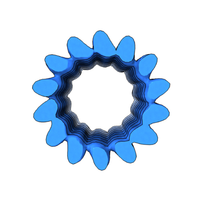 emd_15535.png emd_15535.png | 83.8 KB | ||
| Filedesc metadata | emd-15535.cif.gz | 4.4 KB | ||
| Others | emd_15535_half_map_1.map.gzemd_15535_half_map_2.map.gz | 1.8 MB 1.8 MB | ||
| Archive directory |  http://ftp.pdbj.org/pub/emdb/structures/EMD-15535ftp://ftp.pdbj.org/pub/emdb/structures/EMD-15535 http://ftp.pdbj.org/pub/emdb/structures/EMD-15535ftp://ftp.pdbj.org/pub/emdb/structures/EMD-15535 | HTTPS FTP |
-Related structure data

-Links
| EMDB pages | EMDB (EBI/PDBe) / EMDataResource |
|---|
-Map
| File | Download / File: emd_15535.map.gz / Format: CCP4 / Size: 2 MB / Type: IMAGE STORED AS FLOATING POINT NUMBER (4 BYTES) | ||||||||||||||||||||||||||||||||||||
|---|---|---|---|---|---|---|---|---|---|---|---|---|---|---|---|---|---|---|---|---|---|---|---|---|---|---|---|---|---|---|---|---|---|---|---|---|---|
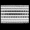
| Annotation | Plasmodium falciparum Gametocyte subpellicular microtubule with 14 protofilaments | ||||||||||||||||||||||||||||||||||||
| Projections & slices | Image control
Images are generated by Spider. | ||||||||||||||||||||||||||||||||||||
| Voxel size | X=Y=Z: 6.712 Å | ||||||||||||||||||||||||||||||||||||
| Density |
| ||||||||||||||||||||||||||||||||||||
| Symmetry | Space group: 1 | ||||||||||||||||||||||||||||||||||||
| Details | EMDB XML:
|
 Z (Sec.)
 Y (Row.)
 X (Col.)


-Supplemental data
-Half map: Unsharpened half map 2
| File | emd_15535_half_map_1.map | ||||||||||||
|---|---|---|---|---|---|---|---|---|---|---|---|---|---|
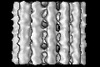
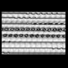
| Annotation | Unsharpened half map 2 | ||||||||||||
| Projections & Slices |
| ||||||||||||
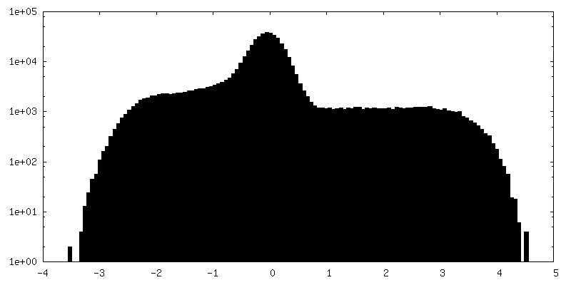
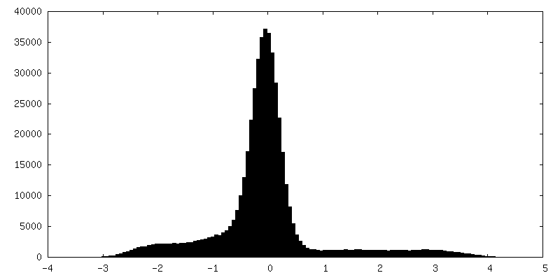
| Density Histograms |


-Half map: Unsharpened half map 1
| File | emd_15535_half_map_2.map | ||||||||||||
|---|---|---|---|---|---|---|---|---|---|---|---|---|---|
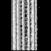
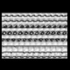
| Annotation | Unsharpened half map 1 | ||||||||||||
| Projections & Slices |
| ||||||||||||
| Density Histograms |


- Sample components
-Entire : Plasmodium falciparum gametocyte
| Entire | Name: Plasmodium falciparum gametocyte |
|---|---|
| Components |
|
-Supramolecule #1: Plasmodium falciparum gametocyte
| Supramolecule | Name: Plasmodium falciparum gametocyte / type: cell / ID: 1 / Parent: 0 / Details: Subpellicular microtubules |
|---|---|
| Source (natural) | Organism: |
-Experimental details
-Structure determination
| Method | cryo EM |
|---|---|
 Processing Processing | subtomogram averaging |
| Aggregation state | cell |
-Sample preparation
| Buffer | pH: 7 / Details: RPMI |
|---|---|
| Grid | Model: UltrAuFoil R1.2/1.3 / Material: GOLD / Mesh: 300 / Support film - Material: GOLD / Support film - topology: HOLEY / Pretreatment - Type: GLOW DISCHARGE / Pretreatment - Time: 60 sec. / Pretreatment - Atmosphere: AIR |
| Vitrification | Cryogen name: ETHANE-PROPANE / Instrument: HOMEMADE PLUNGER |
| Details | FIB-milled Plasmodium falciparum gametocytes |
- Electron microscopy
| Microscope | TFS KRIOS |
|---|---|
| Specialist optics | Energy filter - Name: GIF Bioquantum / Energy filter - Slit width: 20 eV |
| Image recording | Film or detector model: GATAN K3 (6k x 4k) / Average electron dose: 3.0 e/Å2 |
| Electron beam | Acceleration voltage: 300 kV / Electron source:  FIELD EMISSION GUN FIELD EMISSION GUN |
| Electron optics | C2 aperture diameter: 100.0 µm / Illumination mode: FLOOD BEAM / Imaging mode: BRIGHT FIELD / Cs: 2.7 mm / Nominal defocus max: 8.0 µm / Nominal defocus min: 3.0 µm / Nominal magnification: 26000 |
| Sample stage | Specimen holder model: FEI TITAN KRIOS AUTOGRID HOLDER / Cooling holder cryogen: NITROGEN |
| Experimental equipment |  Model: Titan Krios / Image courtesy: FEI Company |
-Image processing
| Final reconstruction | Applied symmetry - Helical parameters - Δz: 8.7 Å Applied symmetry - Helical parameters - Δ&Phi: 25.77 ° Applied symmetry - Helical parameters - Axial symmetry: C14 (14 fold cyclic) Algorithm: BACK PROJECTION / Resolution.type: BY AUTHOR / Resolution: 24.0 Å / Resolution method: FSC 0.143 CUT-OFF / Software - Name: PEET / Number subtomograms used: 11803 |
|---|---|
| Extraction | Number tomograms: 10 / Number images used: 11806 |
| Final angle assignment | Type: OTHER |
| FSC plot (resolution estimation) | 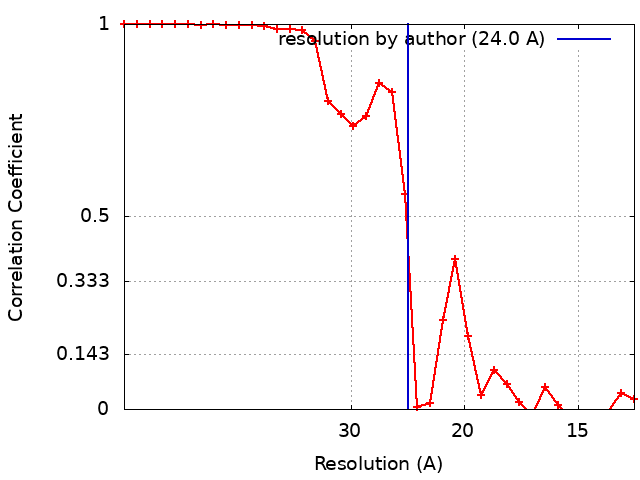 |
