 ムービー
ムービー コントローラー
 構造ビューア
 EMN検索について


-検索条件
-検索結果
検索 (著者・登録者: leis & a)の結果68件中、1から50件目までを表示しています
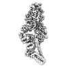
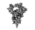
EMDB-37260: 
Cryo-EM structure of Myosin VI in the autoinhibited state (without SAH-extension density)
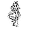
EMDB-37261:
Cryo-EM map of Myosin VI in the autoinhibited state (with SAH-extension density)
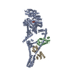
PDB-8w41:
Cryo-EM structure of Myosin VI in the autoinhibited state

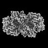
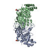
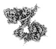
EMDB-41805:
Cryo-EM structure of murine Thrombopoietin receptor ectodomain in complex with Tpo

PDB-8u18:
Cryo-EM structure of murine Thrombopoietin receptor ectodomain in complex with Tpo
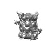
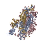
EMDB-42804:
Structure of nucleotide-free Pediculus humanus (Ph) PINK1 dimer

EMDB-42806:
Structure of AMP-PNP-bound Pediculus humanus (Ph) PINK1 dimer

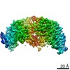
EMDB-42807:
Structure of ADP-bound and phosphorylated Pediculus humanus (Ph) PINK1 dimer
PDB-8uyf:
Structure of nucleotide-free Pediculus humanus (Ph) PINK1 dimer

PDB-8uyh:
Structure of AMP-PNP-bound Pediculus humanus (Ph) PINK1 dimer

PDB-8uyi:
Structure of ADP-bound and phosphorylated Pediculus humanus (Ph) PINK1 dimer

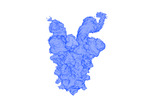
EMDB-27641:
The structure of the interleukin 11 signalling complex, truncated gp130

EMDB-27642:
The structure of the IL-11 signalling complex, with full-length extracellular gp130

PDB-8dps:
The structure of the interleukin 11 signalling complex, truncated gp130

PDB-8dpt:
The structure of the IL-11 signalling complex, with full-length extracellular gp130

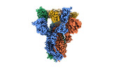
EMDB-40272:
BtCoV-422 in complex with neutralizing antibody JC57-11

EMDB-40306:
BtCoV-422 spike in complex with JC57-11 Fab, global map

EMDB-40310:
BtCoV-422 spike in complex with JC57-11 Fab, local map

PDB-8sak:
BtCoV-422 in complex with neutralizing antibody JC57-11

EMDB-18301:
In-tissue cryo electron tomograms of App^NL-G-F amyloid plaques

EMDB-16018:
Sarkosyl-extracted AppNL-G-F Abeta42 fibril structure

EMDB-16019:
Sarkosyl-extracted AppNL-G-F Abeta42 fibril structure (Methoxy-X04-labelled mice)

PDB-8bfa:
Sarkosyl-extracted AppNL-G-F Abeta42 fibril structure

PDB-8bfb:
Sarkosyl-extracted AppNL-G-F Abeta42 fibril structure (Methoxy-X04-labelled mice)
EMDB-17118:
20S proteasome & CBR3 complex

EMDB-27070:
apo form Cryo-EM structure of Campylobacter jejune ketol-acid reductoisommerase crosslinked by Glutaraldehyde

EMDB-28189:
SARS-CoV-2 Spike in complex with biparatopic nanobody BP10

EMDB-28190:
SARS-CoV-2 RBD in complex with biparatopic nanobody BP10 local refinement

EMDB-14582:
Human heparan sulfate polymerase complex EXT1-EXT2

PDB-7zay:
Human heparan sulfate polymerase complex EXT1-EXT2
EMDB-25618:
SARS-CoV-2 VFLIP spike boung to 2 Ab12 Fab fragments
EMDB-25663:
SARS-CoV-2 S (Spike Glycoprotein) D614G with Three (3) RBDs Up, Bound to Antibody 2-7 scFv, composite map
EMDB-25689:
SARS-CoV-2 S (Spike Glycoprotein) D614G with Three (3) RBDs Up, Bound to Antibody 2-7 scFv, global map with poorly-resolved RBDs and scFvs
EMDB-25690:
SARS-CoV-2 S (Spike Glycoprotein) D614G with Three (3) RBDs Up, Bound to Antibody 2-7 scFv, local refinement map

EMDB-25711:
SARS-CoV-2 S (Spike Glycoprotein) D614G with One(1) RBD Up

PDB-7t3m:
SARS-CoV-2 S (Spike Glycoprotein) D614G with Three (3) RBDs Up, Bound to Antibody 2-7 scFv, composite map
PDB-7t67:
SARS-CoV-2 S (Spike Glycoprotein) D614G with One(1) RBD Up

EMDB-25677:
Structure of dimeric phosphorylated Pediculus humanus (Ph) PINK1
EMDB-25678:
Structure of dimeric phosphorylated Pediculus humanus (Ph) PINK1 with kinked alpha-C helix in chain B

EMDB-25679:
Structure of dimeric phosphorylated Pediculus humanus (Ph) PINK1 with extended alpha-C helix in chain B

EMDB-25680:
Structure of dodecameric unphosphorylated Pediculus humanus (Ph) PINK1 D357A mutant

EMDB-25681:
Structure of dimeric unphosphorylated Pediculus humanus (Ph) PINK1 D357A mutant

PDB-7t4k:
Structure of dimeric phosphorylated Pediculus humanus (Ph) PINK1 with kinked alpha-C helix in chain B

PDB-7t4l:
Structure of dimeric phosphorylated Pediculus humanus (Ph) PINK1 with extended alpha-C helix in chain B

PDB-7t4m:
Structure of dodecameric unphosphorylated Pediculus humanus (Ph) PINK1 D357A mutant

PDB-7t4n:
Structure of dimeric unphosphorylated Pediculus humanus (Ph) PINK1 D357A mutant

EMDB-13662:
Structure of the membrane soluble spike complex from the Lassa virus in a C3-symmetric map

EMDB-13667:
Structure of the membrane soluble spike complex from the Lassa virus in a C1-symmetric map focused on the ectodomain
PDB-7puy:
Structure of the membrane soluble spike complex from the Lassa virus in a C3-symmetric map

PDB-7pvd:
Structure of the membrane soluble spike complex from the Lassa virus in a C1-symmetric map focused on the ectodomain
ページ:
 wwPDBはEMDBデータモデルのバージョン3へ移行します
