 Movie
Movie Controller
 Structure viewers
 About EMN search


-Search query
-Search result
Showing all 37 items for (author: khanna & k)

EMDB-44416: 
Binned tomogram of stationary phase Burkholderia thailandensis
Method: electron tomography / : Khanna K, Welch MD

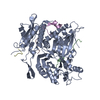
EMDB-25183:
P. chlororaphis 70S ribosome in situ subtomogram average
Method: subtomogram averaging / : Laughlin TG, Deep A, Prichard AM, Seitz C, Gu Y, Enustun E, Suslov S, Khanna K, Birkholz EA, Amaro RE, Pogliano J, Corbett KD, Villa E
EMDB-25220:
In situ subtomogram average of the 201phi2-1 phage nucleus major shell protein, chimallin (concave class)
Method: subtomogram averaging / : Laughlin TG, Deep A, Prichard AM, Seitz C, Gu Y, Enustun E, Suslov S, Khanna K, Birkholz EA, Amaro RE, Pogliano J, Corbett KD, Villa E
EMDB-25221:
In situ consensus subtomogram average of the 201phi2-1 chimallin
Method: subtomogram averaging / : Laughlin TG, Deep A, Prichard AM, Seitz C, Gu Y, Enustun E, Suslov S, Khanna K, Birkholz EA, Amaro RE, Pogliano J, Corbett KD, Villa E
EMDB-25222:
In situ subtomogram average of 201phi2-1 phage nucleus major shell protein, chimallin (intermediate/flat class)
Method: subtomogram averaging / : Laughlin TG, Deep A, Prichard AM, Seitz C, Gu Y, Enustun E, Suslov S, Khanna K, Birkholz EA, Amaro RE, Pogliano J, Corbett KD, Villa E

EMDB-25223:
In situ subtomogram average of the 201phi2-1 phage nucleus major shell protein, chimallin (convex class)
Method: subtomogram averaging / : Laughlin TG, Deep A, Prichard AM, Seitz C, Gu Y, Enustun E, Suslov S, Khanna K, Birkholz EA, Amaro RE, Pogliano J, Corbett KD, Villa E

EMDB-25229:
In situ subtomogram average of the Goslar major phage nucleus shell protein, chimallin (consensus class)
Method: subtomogram averaging / : Laughlin TG, Deep A, Prichard AM, Gu Y, Enustun E, Suslov S, Khanna K, Birkholz EA, Amaro RE, Pogliano J, Corbett KD, Villa E
EMDB-25262:
In situ subtomogram average of Goslar phage nucleus major shell protein, chimallin (concave class)
Method: subtomogram averaging / : Laughlin TG, Deep A, Prichard AM, Seitz C, Gu Y, Enustun E, Suslov S, Khanna K, Birkholz EA, Amaro RE, Pogliano J, Corbett KD, Villa E

EMDB-25358:
In situ subtomogram average of the major Goslar phage nucleus shell protein, chimallin (convex class)
Method: subtomogram averaging / : Laughlin TG, Deep A, Prichard AM, Seitz C, Gu Y, Enustun E, Suslov S, Khanna K, Birkholz EA, Amaro RE, Pogliano J, Corbett KD, Villa E
EMDB-25359:
In situ subtomogram average of the APEC2248 70S ribosome
Method: subtomogram averaging / : Laughlin TG, Deep A, Prichard AM, Seitz C, Gu Y, Enustun E, Suslov S, Khanna K, Birkholz EA, Amaro RE, Pogliano J, Corbett KD, Villa E

EMDB-25360:
In situ subtomogram average of the APEC2248 50S ribosome
Method: subtomogram averaging / : Laughlin TG, Deep A, Prichard AM, Seitz C, Gu Y, Enustun E, Suslov S, Khanna K, Birkholz EA, Amaro RE, Pogliano J, Corbett KD, Villa E
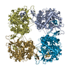
EMDB-25390:
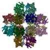
201Phi2-1 Chimallin Cubic (O, 24mer) assembly
Method: single particle / : Laughlin TG, Deep A

EMDB-25391:
201phi2-1 Chimallin localized tetramer reconstruction
Method: single particle / : Laughlin TG, Deep A

EMDB-25392:
201phi2-1 Chimallin C1 localized reconstruction
Method: single particle / : Laughlin TG, Deep A, Prichard AM, Seitz C, Gu Y, Enustun E, Suslov S, Khanna K, Birkholz EA, Amaro RE, Pogliano J, Corbett KD, Villa E

EMDB-25393:
201phi2-1 chimallin rectangular (D4,40mer) assembly
Method: single particle / : Laughlin TG, Deep A, Prichard AM, Seitz C, Gu Y, Enustun E, Suslov S, Khanna K, Birkholz EA, Amaro RE, Pogliano J, Corbett KD, Villa E
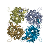
EMDB-25394:
Goslar chimallin cubic (O, 24mer) assembly
Method: single particle / : Laughlin TG, Deep A, Prichard AM, Seitz C, Gu Y, Enustun E, Suslov S, Khanna K, Birkholz EA, Amaro RE, Pogliano J, Corbett KD, Villa E

EMDB-25395:
Goslar chimallin C4 tetramer localized reconstruction
Method: single particle / : Laughlin TG, Deep A, Prichard AM, Seitz C, Gu Y, Enustun E, Suslov S, Khanna K, Birkholz EA, Amaro RE, Pogliano J, Corbett KD, Villa E

EMDB-25396:
Goslar chimallin C1 localized reconstruction
Method: single particle / : Laughlin TG, Deep A, Prichard AM, Seitz C, Gu Y, Enustun E, Suslov S, Khanna K, Birkholz EA, Amaro RE, Pogliano J, Corbett KD, Villa E
PDB-7sqq:
201Phi2-1 Chimallin Cubic (O, 24mer) assembly
Method: single particle / : Laughlin TG, Deep A, Prichard AM, Seitz C, Gu Y, Enustun E, Suslov S, Khanna K, Birkholz EA, Amaro RE, Pogliano J, Corbett KD, Villa E
PDB-7sqr:
201phi2-1 Chimallin localized tetramer reconstruction
Method: single particle / : Laughlin TG, Deep A, Prichard AM, Seitz C, Gu Y, Enustun E, Suslov S, Khanna K, Birkholz EA, Amaro RE, Pogliano J, Corbett KD, Villa E
PDB-7sqs:
201phi2-1 Chimallin C1 localized reconstruction
Method: single particle / : Laughlin TG, Deep A, Prichard AM, Seitz C, Gu Y, Enustun E, Suslov S, Khanna K, Birkholz EA, Amaro RE, Pogliano J, Corbett KD, Villa E
PDB-7sqt:
Goslar chimallin cubic (O, 24mer) assembly
Method: single particle / : Laughlin TG, Deep A, Prichard AM, Seitz C, Gu Y, Enustun E, Suslov S, Khanna K, Birkholz EA, Amaro RE, Pogliano J, Corbett KD, Villa E
PDB-7squ:
Goslar chimallin C4 tetramer localized reconstruction
Method: single particle / : Laughlin TG, Deep A, Prichard AM, Seitz C, Gu Y, Enustun E, Suslov S, Khanna K, Birkholz EA, Amaro RE, Pogliano J, Corbett KD, Villa E
PDB-7sqv:
Goslar chimallin C1 localized reconstruction
Method: single particle / : Laughlin TG, Deep A, Prichard AM, Seitz C, Gu Y, Enustun E, Suslov S, Khanna K, Birkholz EA, Amaro RE, Pogliano J, Corbett KD, Villa E
EMDB-25216:
Slice of a cryo-electron tomogram of PhiPA3-infected Pseudomonas aeruginosa cell at 70 mpi (Cell 4)
Method: electron tomography / : Khanna K, Villa E
EMDB-25217:
Slice of a cryo-electron tomogram of PhiPA3-infected Pseudomonas aeruginosa cell at 70 mpi (Cell 3)
Method: electron tomography / : Khanna K, Villa E
EMDB-25218:
Slice of a cryo-electron tomogram of PhiPA3-infected Pseudomonas aeruginosa cell at 70 mpi (Cell 2)
Method: electron tomography / : Khanna K, Villa E
EMDB-25219:
Slice of a cryo-electron tomogram of PhiPA3-infected Pseudomonas aeruginosa cell at 70 mpi showing phage bouquets (same as Fig 4A of the manuscript)
Method: electron tomography / : Khanna K, Villa E
EMDB-23963:
Tomogram of a dividing vegetative cell of Bacillus subtilis (Figure 3A of the manuscript Khanna et al., 2021)
Method: electron tomography / : Khanna K, Villa E
EMDB-23964:
Tomogram of a dividing vegetative cell of Bacillus subtilis (Figure 4A of the manuscript Khanna et al., 2021)
Method: electron tomography / : Khanna K, Villa E
EMDB-23965:
Tomogram of a dividing vegetative cell of Bacillus subtilis FtsZ-linker(Q-rich) strain (Figure 5A and 6A of the manuscript Khanna et al., 2021)
Method: electron tomography / : Khanna K, Villa E
EMDB-23966:
Tomogram of a dividing sporulating cell of Bacillus subtilis (Figure 7A of the manuscript Khanna et al., 2021)
Method: electron tomography / : Khanna K, Villa E
EMDB-23967:
Tomogram of a dividing sporulating cell of Bacillus subtilis (Figure 7 - figure supplement 4 of the manuscript Khanna et al., 2021)
Method: electron tomography / : Khanna K, Villa E

EMDB-23968:
Tomogram of a dividing sporulating cell of Bacillus subtilis SpoIIE null mutant (Figure 8A of the manuscript Khanna et al., 2001)
Method: electron tomography / : Khanna K, Villa E

EMDB-20335:
Cryo-electron tomogram of Bacillus subtilis wild type sporangium (flat septum) shown in Figure 1D of the publication
Method: electron tomography / : Khanna K, Villa E
EMDB-20336:
Cryo-electron tomogram of Bacillus subtilis wild type sporangium (curved septum) shown in Figure 1F of the publication
Method: electron tomography / : Khanna K, Villa E
EMDB-20337:
Cryo-electron tomogram of Bacillus subtilis wild type sporangium (engulfing septum) shown in Figure 1H of the publication
Method: electron tomography / : Khanna K, Villa E
 wwPDB to switch to version 3 of the EMDB data model
