Movie
Movie Controller

[English] 日本語
 Yorodumi
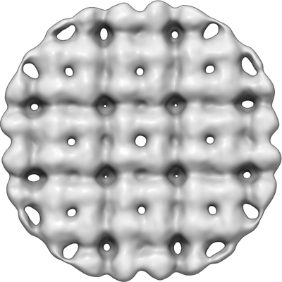
- EMDB-25221: In situ consensus subtomogram average of the 201phi2-1 chimallin -
+ Open data

- Basic information
| Entry |  | ||||||||||||
|---|---|---|---|---|---|---|---|---|---|---|---|---|---|
| Title | In situ consensus subtomogram average of the 201phi2-1 chimallin | ||||||||||||
 Map data Map data | |||||||||||||
 Sample Sample |
| ||||||||||||
| Biological species |  Pseudomonas phage 201phi2-1 (virus) Pseudomonas phage 201phi2-1 (virus) | ||||||||||||
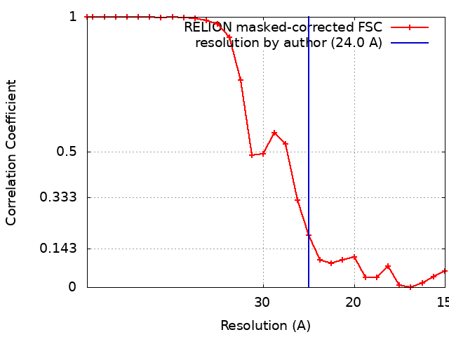
| Method | subtomogram averaging / cryo EM / Resolution: 24.0 Å | ||||||||||||
 Authors Authors | Laughlin TG / Deep A / Prichard AM / Seitz C / Gu Y / Enustun E / Suslov S / Khanna K / Birkholz EA / Amaro RE ...Laughlin TG / Deep A / Prichard AM / Seitz C / Gu Y / Enustun E / Suslov S / Khanna K / Birkholz EA / Amaro RE / Pogliano J / Corbett KD / Villa E | ||||||||||||
| Funding support |  United States, 3 items United States, 3 items
| ||||||||||||
 Citation Citation | Journal: Nature / Year: 2022 Title: Architecture and self-assembly of the jumbo bacteriophage nuclear shell. Authors: Thomas G Laughlin / Amar Deep / Amy M Prichard / Christian Seitz / Yajie Gu / Eray Enustun / Sergey Suslov / Kanika Khanna / Erica A Birkholz / Emily Armbruster / J Andrew McCammon / Rommie ...Authors: Thomas G Laughlin / Amar Deep / Amy M Prichard / Christian Seitz / Yajie Gu / Eray Enustun / Sergey Suslov / Kanika Khanna / Erica A Birkholz / Emily Armbruster / J Andrew McCammon / Rommie E Amaro / Joe Pogliano / Kevin D Corbett / Elizabeth Villa / Abstract: Bacteria encode myriad defences that target the genomes of infecting bacteriophage, including restriction-modification and CRISPR-Cas systems. In response, one family of large bacteriophages uses a ...Bacteria encode myriad defences that target the genomes of infecting bacteriophage, including restriction-modification and CRISPR-Cas systems. In response, one family of large bacteriophages uses a nucleus-like compartment to protect its replicating genomes by excluding host defence factors. However, the principal composition and structure of this compartment remain unknown. Here we find that the bacteriophage nuclear shell assembles primarily from one protein, which we name chimallin (ChmA). Combining cryo-electron tomography of nuclear shells in bacteriophage-infected cells and cryo-electron microscopy of a minimal chimallin compartment in vitro, we show that chimallin self-assembles as a flexible sheet into closed micrometre-scale compartments. The architecture and assembly dynamics of the chimallin shell suggest mechanisms for its nucleation and growth, and its role as a scaffold for phage-encoded factors mediating macromolecular transport, cytoskeletal interactions, and viral maturation. | ||||||||||||
| History |
|
- Structure visualization
| Supplemental images |
|---|
- Downloads & links
-EMDB archive
| Map data | emd_25221.map.gz | 491.6 KB |  EMDB map data format EMDB map data format | |
|---|---|---|---|---|
| Header (meta data) | emd-25221-v30.xmlemd-25221.xml | 17.6 KB 17.6 KB | Display Display | EMDB header |
| FSC (resolution estimation) | emd_25221_fsc.xml | 2.4 KB | Display | FSC data file |
| Images |  emd_25221.png emd_25221.png | 90.6 KB | ||
| Masks | emd_25221_msk_1.map | 1 MB | Mask map | |
| Others | emd_25221_half_map_1.map.gzemd_25221_half_map_2.map.gz | 493.7 KB 494 KB | ||
| Archive directory |  http://ftp.pdbj.org/pub/emdb/structures/EMD-25221ftp://ftp.pdbj.org/pub/emdb/structures/EMD-25221 http://ftp.pdbj.org/pub/emdb/structures/EMD-25221ftp://ftp.pdbj.org/pub/emdb/structures/EMD-25221 | HTTPS FTP |
-Related structure data
| Related structure data |  7sqqC 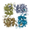 7sqrC 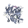 7sqsC 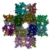 7sqtC 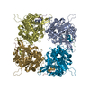 7squC 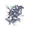 7sqvC C: citing same article ( |
|---|---|
| EM raw data | EMPIAR-10859 (Title: In situ cryo-electron tomography of P. chlororaphis infected by 201phi2-1 Data size: 14.5 Data #1: Unaligned tilt-movie frames as unormalized LZW-TIFF [micrographs - multiframe]) |


-Links
| EMDB pages | EMDB (EBI/PDBe) / EMDataResource |
|---|
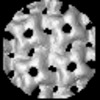
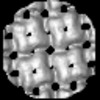
-Map
| File | Download / File: emd_25221.map.gz / Format: CCP4 / Size: 1 MB / Type: IMAGE STORED AS FLOATING POINT NUMBER (4 BYTES) | ||||||||||||||||||||||||||||||||||||
|---|---|---|---|---|---|---|---|---|---|---|---|---|---|---|---|---|---|---|---|---|---|---|---|---|---|---|---|---|---|---|---|---|---|---|---|---|---|
| Projections & slices | Image control
Images are generated by Spider. | ||||||||||||||||||||||||||||||||||||
| Voxel size | X=Y=Z: 7.5 Å | ||||||||||||||||||||||||||||||||||||
| Density |
| ||||||||||||||||||||||||||||||||||||
| Symmetry | Space group: 1 | ||||||||||||||||||||||||||||||||||||
| Details | EMDB XML:
|
 Z (Sec.)
 Y (Row.)
 X (Col.)


-Supplemental data
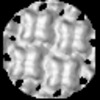
-Mask #1
| File | emd_25221_msk_1.map | ||||||||||||
|---|---|---|---|---|---|---|---|---|---|---|---|---|---|
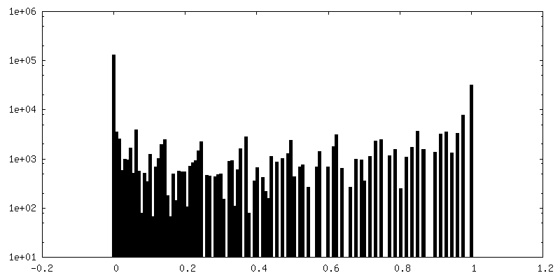
| Projections & Slices |
| ||||||||||||
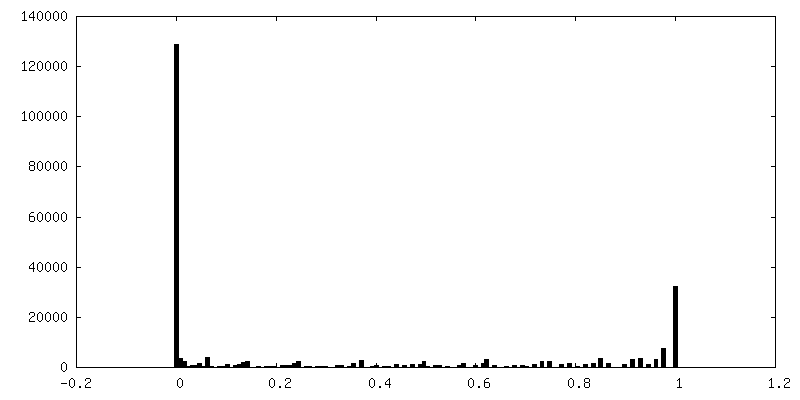
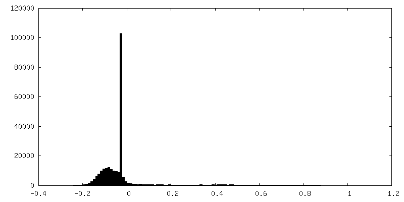
| Density Histograms |


-Half map: #1
| File | emd_25221_half_map_1.map | ||||||||||||
|---|---|---|---|---|---|---|---|---|---|---|---|---|---|
| Projections & Slices |
| ||||||||||||
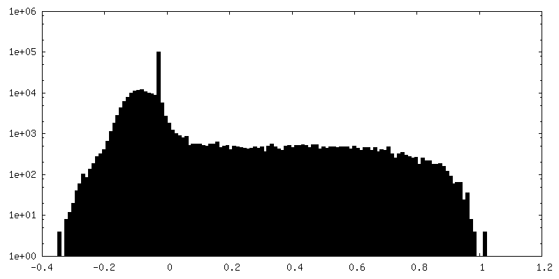
| Density Histograms |


-Half map: #2
| File | emd_25221_half_map_2.map | ||||||||||||
|---|---|---|---|---|---|---|---|---|---|---|---|---|---|
| Projections & Slices |
| ||||||||||||
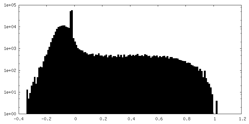
| Density Histograms |


- Sample components
-Entire : 201phi2-1 jumbo phage major shell protein, chimallin
| Entire | Name: 201phi2-1 jumbo phage major shell protein, chimallin |
|---|---|
| Components |
|
-Supramolecule #1: 201phi2-1 jumbo phage major shell protein, chimallin
| Supramolecule | Name: 201phi2-1 jumbo phage major shell protein, chimallin / type: organelle_or_cellular_component / ID: 1 / Parent: 0 |
|---|---|
| Source (natural) | Organism: Pseudomonas phage 201phi2-1 (virus) |
-Experimental details
-Structure determination
| Method | cryo EM |
|---|---|
 Processing Processing | subtomogram averaging |
| Aggregation state | cell |
-Sample preparation
| Buffer | pH: 7 |
|---|---|
| Grid | Model: Quantifoil R2/1 / Material: COPPER / Mesh: 200 / Support film - Material: CARBON / Support film - topology: HOLEY ARRAY / Pretreatment - Type: GLOW DISCHARGE / Pretreatment - Atmosphere: AIR / Pretreatment - Pressure: 0.019 kPa / Details: 20 mA, PELCO EasiGLO |
| Vitrification | Cryogen name: ETHANE-PROPANE / Instrument: HOMEMADE PLUNGER / Details: manual plunger, backside blotted for ~7 seconds. |
- Electron microscopy
| Microscope | FEI TITAN KRIOS |
|---|---|
| Image recording | Film or detector model: GATAN K2 SUMMIT (4k x 4k) / Number grids imaged: 1 / Average electron dose: 1.8 e/Å2 |
| Electron beam | Acceleration voltage: 300 kV / Electron source:  FIELD EMISSION GUN FIELD EMISSION GUN |
| Electron optics | C2 aperture diameter: 70.0 µm / Illumination mode: FLOOD BEAM / Imaging mode: BRIGHT FIELD / Cs: 2.7 mm / Nominal magnification: 42000 |
| Sample stage | Specimen holder model: FEI TITAN KRIOS AUTOGRID HOLDER / Cooling holder cryogen: NITROGEN |
| Experimental equipment |  Model: Titan Krios / Image courtesy: FEI Company |