 Movie
Movie Controller

[English] 日本語
 Yorodumi
- EMDB-23964: Tomogram of a dividing vegetative cell of Bacillus subtilis (Figu... -
+ Open data

- Basic information
| Entry | Database: EMDB / ID: EMD-23964 | |||||||||
|---|---|---|---|---|---|---|---|---|---|---|
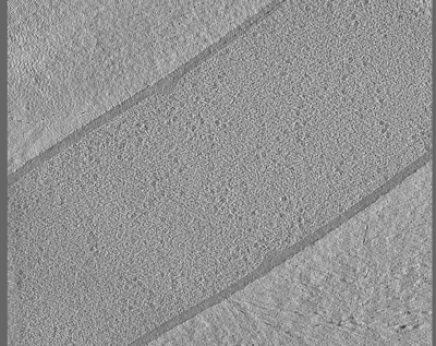
| Title | Tomogram of a dividing vegetative cell of Bacillus subtilis (Figure 4A of the manuscript Khanna et al., 2021) | |||||||||
 Map data Map data | Tomogram of a dividing cell of Bacillus subtilis during vegetative growth (Figure 4A of the manuscript Khanna et al., 2021) | |||||||||
 Sample Sample |
| |||||||||
| Biological species |  | |||||||||
| Method | electron tomography / cryo EM | |||||||||
 Authors Authors | Khanna K / Villa E | |||||||||
| Funding support |  United States, 2 items United States, 2 items
| |||||||||
 Citation Citation | Journal: Elife / Year: 2021 Title: Asymmetric localization of the cell division machinery during sporulation. Authors: Kanika Khanna / Javier Lopez-Garrido / Joseph Sugie / Kit Pogliano / Elizabeth Villa / Abstract: The Gram-positive bacterium can divide via two modes. During vegetative growth, the division septum is formed at the midcell to produce two equal daughter cells. However, during sporulation, the ...The Gram-positive bacterium can divide via two modes. During vegetative growth, the division septum is formed at the midcell to produce two equal daughter cells. However, during sporulation, the division septum is formed closer to one pole to yield a smaller forespore and a larger mother cell. Using cryo-electron tomography, genetics and fluorescence microscopy, we found that the organization of the division machinery is different in the two septa. While FtsAZ filaments, the major orchestrators of bacterial cell division, are present uniformly around the leading edge of the invaginating vegetative septa, they are only present on the mother cell side of the invaginating sporulation septa. We provide evidence suggesting that the different distribution and number of FtsAZ filaments impact septal thickness, causing vegetative septa to be thicker than sporulation septa already during constriction. Finally, we show that a sporulation-specific protein, SpoIIE, regulates asymmetric divisome localization and septal thickness during sporulation. | |||||||||
| History |
|
- Structure visualization
| Movie |
 Movie viewer Movie viewer |
|---|---|
| Supplemental images |
- Downloads & links
-EMDB archive
| Map data | emd_23964.map.gz | 284.2 MB | EMDB map data format | |
|---|---|---|---|---|
| Header (meta data) | emd-23964-v30.xmlemd-23964.xml | 9.2 KB 9.2 KB | Display Display | EMDB header |
| Images |  emd_23964.png emd_23964.png | 189.6 KB | ||
| Archive directory |  http://ftp.pdbj.org/pub/emdb/structures/EMD-23964ftp://ftp.pdbj.org/pub/emdb/structures/EMD-23964 http://ftp.pdbj.org/pub/emdb/structures/EMD-23964ftp://ftp.pdbj.org/pub/emdb/structures/EMD-23964 | HTTPS FTP |
-Related structure data
| Related structure data | C: citing same article ( |
|---|---|
| EM raw data | EMPIAR-10710 (Title: Tilt series of dividing vegetative and sporulating cells of Bacillus subtilis from the manuscript - Khanna et al., 2021 Data size: 8.7 Data #1: Tilt series of a dividing vegetative cell of Bacillus subtilis (Figure 3A of the manuscript Khanna et al., 2021) [tilt series] Data #2: Tomogram of a dividing vegetative cell of Bacillus subtilis (Figure 4A of the manuscript Khanna et al., 2021) [tilt series] Data #3: Tomogram of a dividing vegetative cell of Bacillus subtilis FtsZ-linker(Q-rich) strain (Figure 5A and 6A of the manuscript Khanna et al., 2021) [tilt series] Data #4: Tomogram of a dividing sporulating cell of Bacillus subtilis (Figure 7A of the manuscript Khanna et al., 2021) [tilt series] Data #5: Tomogram of a dividing sporulating cell of Bacillus subtilis (Figure 7 - figure supplement 4 of the manuscript Khanna et al., 2021) [tilt series] Data #6: Tomogram of a dividing sporulating cell of Bacillus subtilis SpoIIE null mutant (Figure 8A of the manuscript Khanna et al., 2001) [tilt series]) |

-Links
| EMDB pages | EMDB (EBI/PDBe) / EMDataResource |
|---|
-Map
| File | Download / File: emd_23964.map.gz / Format: CCP4 / Size: 424.8 MB / Type: IMAGE STORED AS SIGNED BYTE | ||||||||||||||||||||||||||||||||||||||||||||||||||||||||||||||||||||
|---|---|---|---|---|---|---|---|---|---|---|---|---|---|---|---|---|---|---|---|---|---|---|---|---|---|---|---|---|---|---|---|---|---|---|---|---|---|---|---|---|---|---|---|---|---|---|---|---|---|---|---|---|---|---|---|---|---|---|---|---|---|---|---|---|---|---|---|---|---|
| Annotation | Tomogram of a dividing cell of Bacillus subtilis during vegetative growth (Figure 4A of the manuscript Khanna et al., 2021) | ||||||||||||||||||||||||||||||||||||||||||||||||||||||||||||||||||||
| Voxel size | X=Y=Z: 17.06 Å | ||||||||||||||||||||||||||||||||||||||||||||||||||||||||||||||||||||
| Density |
| ||||||||||||||||||||||||||||||||||||||||||||||||||||||||||||||||||||
| Symmetry | Space group: 1 | ||||||||||||||||||||||||||||||||||||||||||||||||||||||||||||||||||||
| Details | EMDB XML:
CCP4 map header:
| ||||||||||||||||||||||||||||||||||||||||||||||||||||||||||||||||||||
-Supplemental data
- Sample components
-Entire : Dividing vegetative cell of Bacillus subtilis
| Entire | Name: Dividing vegetative cell of Bacillus subtilis |
|---|---|
| Components |
|
-Supramolecule #1: Dividing vegetative cell of Bacillus subtilis
| Supramolecule | Name: Dividing vegetative cell of Bacillus subtilis / type: cell / ID: 1 / Parent: 0 / Details: Figure 4A of the manuscript Khanna et al., 2021 |
|---|---|
| Source (natural) | Organism: |
-Experimental details
-Structure determination
| Method | cryo EM |
|---|---|
 Processing Processing | electron tomography |
| Aggregation state | cell |
-Sample preparation
| Buffer | pH: 7 |
|---|---|
| Vitrification | Cryogen name: ETHANE-PROPANE |
| Sectioning | Focused ion beam - Instrument: OTHER / Focused ion beam - Ion: OTHER / Focused ion beam - Voltage: 5 kV / Focused ion beam - Current: 0.05 nA / Focused ion beam - Duration: 1800 sec. / Focused ion beam - Temperature: 93 K / Focused ion beam - Initial thickness: 1200 nm / Focused ion beam - Final thickness: 200 nm Focused ion beam - Details: The value given for _emd_sectioning_focused_ion_beam.instrument is Thermo Scientific Aquilos. This is not in a list of allowed values {'DB235', 'OTHER'} so OTHER is written into the XML file. |
- Electron microscopy
| Microscope | FEI TITAN KRIOS |
|---|---|
| Image recording | Film or detector model: GATAN K2 SUMMIT (4k x 4k) / Average electron dose: 1.0 e/Å2 |
| Electron beam | Acceleration voltage: 300 kV / Electron source:  FIELD EMISSION GUN FIELD EMISSION GUN |
| Electron optics | Illumination mode: FLOOD BEAM / Imaging mode: BRIGHT FIELD |
| Experimental equipment |  Model: Titan Krios / Image courtesy: FEI Company |
-Image processing
| Final reconstruction | Software - Name: IMOD / Number images used: 48 |
|---|
