National Institutes of Health/National Institute of General Medical Sciences (NIH/NIGMS)
GM104556
United States
National Institutes of Health/National Institute of General Medical Sciences (NIH/NIGMS)
GM129245
United States
National Science Foundation (NSF, United States)
DBI 1920374
United States
Citation
Journal: Sci Adv / Year: 2022 Title: Subcellular organization of viral particles during maturation of nucleus-forming jumbo phage. Authors: Vorrapon Chaikeeratisak / Kanika Khanna / Katrina T Nguyen / MacKennon E Egan / Eray Enustun / Emily Armbruster / Jina Lee / Kit Pogliano / Elizabeth Villa / Joe Pogliano / Abstract: Many eukaryotic viruses assemble mature particles within distinct subcellular compartments, but bacteriophages are generally assumed to assemble randomly throughout the host cell cytoplasm. Here, we ...Many eukaryotic viruses assemble mature particles within distinct subcellular compartments, but bacteriophages are generally assumed to assemble randomly throughout the host cell cytoplasm. Here, we show that viral particles of nucleus-forming jumbo phage PhiPA3 assemble into a unique structure inside cells we term phage bouquets. We show that after capsids complete DNA packaging at the surface of the phage nucleus, tails assemble and attach to capsids, and these particles accumulate over time in a spherical pattern, with tails oriented inward and the heads outward to form bouquets at specific subcellular locations. Bouquets localize at the same fixed distance from the phage nucleus even when it is mispositioned, suggesting an active mechanism for positioning. These results mark the discovery of a pathway for organizing mature viral particles inside bacteria and demonstrate that nucleus-forming jumbo phages, like most eukaryotic viruses, are highly spatially organized during all stages of their lytic cycle.
Download / File: emd_25217.map.gz / Format: CCP4 / Size: 531.1 MB / Type: IMAGE STORED AS SIGNED BYTE
Annotation
Slice of a cryo-electron tomogram of PhiPA3-infected Pseudomonas aeruginosa cell at 70 mpi (Cell 3)
Voxel size
X=Y=Z: 22.46 Å
Density
Minimum - Maximum
-128.0 - 127.0
Average (Standard dev.)
32.027630000000002 (±8.504201)
Symmetry
Space group: 1
Details
EMDB XML:
Map geometry
Axis order
X
Y
Z
Origin
0
0
176
Dimensions
1280
1236
352
Spacing
1236
1280
352
Cell
A: 27760.559 Å / B: 28748.799 Å / C: 7905.92 Å α=β=γ: 90.0 °
-
Supplemental data
-
Sample components
-
Entire : Pseudomonas aeruginosa infected with bacteriophage PhiPA3 at 75 mpi
Entire
Name: Pseudomonas aeruginosa infected with bacteriophage PhiPA3 at 75 mpi
Components
Cell: Pseudomonas aeruginosa infected with bacteriophage PhiPA3 at 75 mpi
-
Supramolecule #1: Pseudomonas aeruginosa infected with bacteriophage PhiPA3 at 75 mpi
Supramolecule
Name: Pseudomonas aeruginosa infected with bacteriophage PhiPA3 at 75 mpi type: cell / ID: 1 / Parent: 0
Source (natural)
Organism: Pseudomonas aeruginosa PA1 (bacteria)
-
Experimental details
-
Structure determination
Method
cryo EM
Processing
electron tomography
Aggregation state
cell
-
Sample preparation
Buffer
pH: 7
Vitrification
Cryogen name: ETHANE-PROPANE
Sectioning
Focused ion beam - Instrument: OTHER / Focused ion beam - Ion: OTHER / Focused ion beam - Voltage: 30 / Focused ion beam - Current: 0.03 / Focused ion beam - Duration: 1800 / Focused ion beam - Temperature: 93 K / Focused ion beam - Initial thickness: 2000 / Focused ion beam - Final thickness: 150 Focused ion beam - Details: The value given for _em_focused_ion_beam.instrument is FEI Scios. This is not in a list of allowed values {'DB235', 'OTHER'} so OTHER is written into the XML file.
-
Electron microscopy
Microscope
FEI POLARA 300
Image recording
Film or detector model: GATAN K2 SUMMIT (4k x 4k) / Average electron dose: 1.0 e/Å2
Electron beam
Acceleration voltage: 300 kV / Electron source: FIELD EMISSION GUN
Electron optics
Illumination mode: OTHER / Imaging mode: BRIGHT FIELD
Experimental equipment
Model: Tecnai Polara / Image courtesy: FEI Company
-
Image processing
Final reconstruction
Number images used: 60
+
About Yorodumi
-
News
-
Feb 9, 2022. New format data for meta-information of EMDB entries
New format data for meta-information of EMDB entries
Version 3 of the EMDB header file is now the official format.
The previous official version 1.9 will be removed from the archive.
In the structure databanks used in Yorodumi, some data are registered as the other names, "COVID-19 virus" and "2019-nCoV". Here are the details of the virus and the list of structure data.
Jan 31, 2019. EMDB accession codes are about to change! (news from PDBe EMDB page)
EMDB accession codes are about to change! (news from PDBe EMDB page)
The allocation of 4 digits for EMDB accession codes will soon come to an end. Whilst these codes will remain in use, new EMDB accession codes will include an additional digit and will expand incrementally as the available range of codes is exhausted. The current 4-digit format prefixed with “EMD-” (i.e. EMD-XXXX) will advance to a 5-digit format (i.e. EMD-XXXXX), and so on. It is currently estimated that the 4-digit codes will be depleted around Spring 2019, at which point the 5-digit format will come into force.
The EM Navigator/Yorodumi systems omit the EMD- prefix.
Related info.:Q: What is EMD? / ID/Accession-code notation in Yorodumi/EM Navigator
Yorodumi is a browser for structure data from EMDB, PDB, SASBDB, etc.
This page is also the successor to EM Navigator detail page, and also detail information page/front-end page for Omokage search.
The word "yorodu" (or yorozu) is an old Japanese word meaning "ten thousand". "mi" (miru) is to see.
Related info.:EMDB / PDB / SASBDB / Comparison of 3 databanks / Yorodumi Search / Aug 31, 2016. New EM Navigator & Yorodumi / Yorodumi Papers / Jmol/JSmol / Function and homology information / Changes in new EM Navigator and Yorodumi
 Movie
Movie Controller

 Yorodumi
 Open data
 Basic information
 Map data
 Sample
 Keywords
 Pseudomonas aeruginosa PA1 (bacteria)
 Authors
 United States, 3 items
  Citation
 Structure visualization
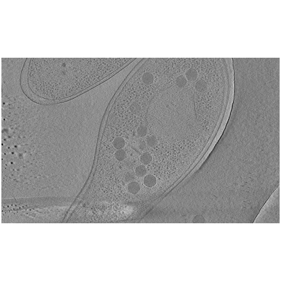
 Downloads & links
 EMDB map data format
 emd_25217.png
 http://ftp.pdbj.org/pub/emdb/structures/EMD-25217
 Sample components
 Processing
 Electron microscopy
 FIELD EMISSION GUN
