National Institutes of Health/National Institute of General Medical Sciences (NIH/NIGMS)
GM104556
米国
National Institutes of Health/National Institute of General Medical Sciences (NIH/NIGMS)
GM129245
米国
National Science Foundation (NSF, United States)
DBI 1920374
米国
引用
ジャーナル: Sci Adv / 年: 2022 タイトル: Subcellular organization of viral particles during maturation of nucleus-forming jumbo phage. 著者: Vorrapon Chaikeeratisak / Kanika Khanna / Katrina T Nguyen / MacKennon E Egan / Eray Enustun / Emily Armbruster / Jina Lee / Kit Pogliano / Elizabeth Villa / Joe Pogliano / 要旨: Many eukaryotic viruses assemble mature particles within distinct subcellular compartments, but bacteriophages are generally assumed to assemble randomly throughout the host cell cytoplasm. Here, we ...Many eukaryotic viruses assemble mature particles within distinct subcellular compartments, but bacteriophages are generally assumed to assemble randomly throughout the host cell cytoplasm. Here, we show that viral particles of nucleus-forming jumbo phage PhiPA3 assemble into a unique structure inside cells we term phage bouquets. We show that after capsids complete DNA packaging at the surface of the phage nucleus, tails assemble and attach to capsids, and these particles accumulate over time in a spherical pattern, with tails oriented inward and the heads outward to form bouquets at specific subcellular locations. Bouquets localize at the same fixed distance from the phage nucleus even when it is mispositioned, suggesting an active mechanism for positioning. These results mark the discovery of a pathway for organizing mature viral particles inside bacteria and demonstrate that nucleus-forming jumbo phages, like most eukaryotic viruses, are highly spatially organized during all stages of their lytic cycle.
ダウンロード / ファイル: emd_25217.map.gz / 形式: CCP4 / 大きさ: 531.1 MB / タイプ: IMAGE STORED AS SIGNED BYTE
注釈
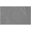
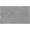
Slice of a cryo-electron tomogram of PhiPA3-infected Pseudomonas aeruginosa cell at 70 mpi (Cell 3)
ボクセルのサイズ
X=Y=Z: 22.46 Å
密度
最小 - 最大
-128.0 - 127.0
平均 (標準偏差)
32.027630000000002 (±8.504201)
対称性
空間群: 1
詳細
EMDB XML:
マップ形状
Axis order
X
Y
Z
Origin
0
0
176
サイズ
1280
1236
352
Spacing
1236
1280
352
セル
A: 27760.559 Å / B: 28748.799 Å / C: 7905.92 Å α=β=γ: 90.0 °
-
添付データ
-
試料の構成要素
-
全体 : Pseudomonas aeruginosa infected with bacteriophage PhiPA3 at 75 mpi
全体
名称: Pseudomonas aeruginosa infected with bacteriophage PhiPA3 at 75 mpi
要素
細胞: Pseudomonas aeruginosa infected with bacteriophage PhiPA3 at 75 mpi
-
超分子 #1: Pseudomonas aeruginosa infected with bacteriophage PhiPA3 at 75 mpi
超分子
名称: Pseudomonas aeruginosa infected with bacteriophage PhiPA3 at 75 mpi タイプ: cell / ID: 1 / 親要素: 0
由来(天然)
生物種: Pseudomonas aeruginosa PA1 (緑膿菌)
-
実験情報
-
構造解析
手法
クライオ電子顕微鏡法
解析
電子線トモグラフィー法
試料の集合状態
cell
-
試料調製
緩衝液
pH: 7
凍結
凍結剤: ETHANE-PROPANE
切片作成
集束イオンビーム - 装置: OTHER / 集束イオンビーム - イオン: OTHER / 集束イオンビーム - 電圧: 30 / 集束イオンビーム - 電流: 0.03 / 集束イオンビーム - 時間: 1800 / 集束イオンビーム - 温度: 93 K / 集束イオンビーム - Initial thickness: 2000 / 集束イオンビーム - 最終 厚さ: 150 集束イオンビーム - 詳細: The value given for _em_focused_ion_beam.instrument is FEI Scios. This is not in a list of allowed values {'DB235', 'OTHER'} so OTHER is written into the XML file.
-
電子顕微鏡法
顕微鏡
FEI POLARA 300
電子線
加速電圧: 300 kV / 電子線源: FIELD EMISSION GUN
電子光学系
照射モード: OTHER / 撮影モード: BRIGHT FIELDBright-field microscopy
撮影
フィルム・検出器のモデル: GATAN K2 SUMMIT (4k x 4k) 平均電子線量: 1.0 e/Å2
 ムービー
ムービー コントローラー


 データを開く
 基本情報
基本情報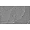
 マップデータ
 試料
試料 キーワード
キーワード Bacteriophage (ファージ) / Phage Bouquet / Jumbo phage infection /
 データ登録者
 米国, 3件
  引用
引用
 構造の表示
構造の表示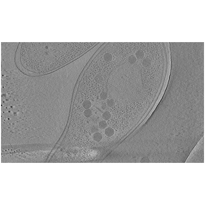
 ダウンロードとリンク
 EMDBマップデータ形式
 emd_25217.png
 http://ftp.pdbj.org/pub/emdb/structures/EMD-25217
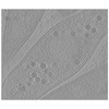
 試料の構成要素
 解析
解析 電子顕微鏡法
