 ムービー
ムービー コントローラー
 構造ビューア
 EMN検索について


-検索条件
-検索結果
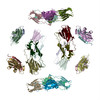
検索 (著者・登録者: gowen & b)の結果全20件を表示しています
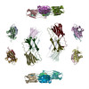
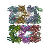
PDB-2h50: 
Multiple distinct assemblies reveal conformational flexibility in the small heat shock protein Hsp26
PDB-2h53:
Multiple distinct assemblies reveal conformational flexibility in the small heat shock protein Hsp26
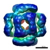
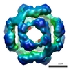
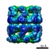
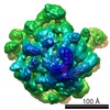
EMDB-1221:
Multiple distinct assemblies reveal conformational flexibility in the small heat shock protein Hsp26.
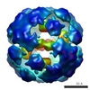
EMDB-1226:
Multiple distinct assemblies reveal conformational flexibility in the small heat shock protein Hsp26.
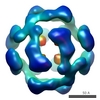
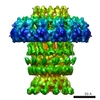
EMDB-1227:
Multiple distinct assemblies reveal conformational flexibility in the small heat shock protein Hsp26.
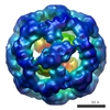
EMDB-1228:
Multiple distinct assemblies reveal conformational flexibility in the small heat shock protein Hsp26.
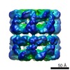
EMDB-1229:
Multiple distinct assemblies reveal conformational flexibility in the small heat shock protein Hsp26.
EMDB-1230:
Multiple distinct assemblies reveal conformational flexibility in the small heat shock protein Hsp26.
PDB-2c7e:
REVISED ATOMIC STRUCTURE FITTING INTO A GROEL(D398A)-ATP7 CRYO-EM MAP (EMD 1047)

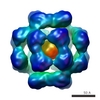
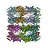
EMDB-1046:
ATP-bound states of GroEL captured by cryo-electron microscopy.
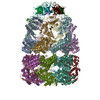
EMDB-1047:
ATP-bound states of GroEL captured by cryo-electron microscopy.

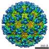
EMDB-1020:
Structure of a viral DNA gatekeeper at 10 A resolution by cryo-electron microscopy.
EMDB-1021:
Structure of a viral DNA gatekeeper at 10 A resolution by cryo-electron microscopy.
EMDB-1042:
ATP-bound states of GroEL captured by cryo-electron microscopy.

EMDB-1018:
The first step: activation of the Semliki Forest virus spike protein precursor causes a localized conformational change in the trimeric spike.
EMDB-1019:
The Escherichia coli large ribosomal subunit at 7.5 A resolution.
EMDB-1015:
Cryo-electron microscopy reveals the functional organization of an enveloped virus, Semliki Forest virus.
PDB-1gr5:
Solution Structure of apo GroEL by Cryo-Electron microscopy
PDB-1gru:
SOLUTION STRUCTURE OF GROES-ADP7-GROEL-ATP7 COMPLEX BY CRYO-EM
PDB-1dyl:
9 ANGSTROM RESOLUTION CRYO-EM RECONSTRUCTION STRUCTURE OF SEMLIKI FOREST VIRUS (SFV) AND FITTING OF THE CAPSID PROTEIN STRUCTURE IN THE EM DENSITY
 wwPDBはEMDBデータモデルのバージョン3へ移行します
