 Movie
Movie Controller
 Structure viewers
 About EMN search


-Search query
-Search result
Showing 1 - 50 of 71 items for (author: dickinson & s)

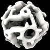
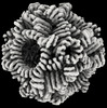
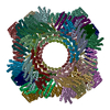
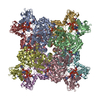
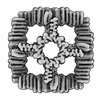
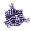
EMDB-41907: 
Computationally Designed, Expandable O4 Octahedral Handshake Nanocage
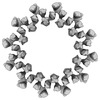
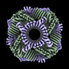
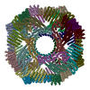
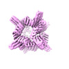
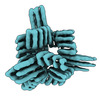
EMDB-42031:
Computational Designed Nanocage O43_129_+8
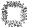
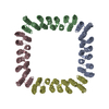
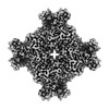
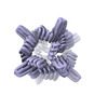
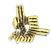
EMDB-43318:
Twistless helix 12 repeat ring design R12B
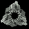
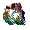
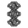
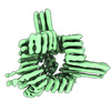
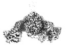
EMDB-29974:
Cryo-EM structure of synthetic tetrameric building block sC4
EMDB-41364:
CryoEM Structure of a Computationally Designed T3 Tetrahedral Nanocage
EMDB-42906:
Computational Designed Nanocage O43_129
EMDB-42944:
Computational Designed Nanocage O43_129_+4
PDB-8gel:
Cryo-EM structure of synthetic tetrameric building block sC4
PDB-8tl7:
CryoEM Structure of a Computationally Designed T3 Tetrahedral Nanocage
PDB-8v2d:
Computational Designed Nanocage O43_129
PDB-8v3b:
Computational Designed Nanocage O43_129_+4
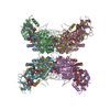
EMDB-41986:
Human retinal variant phosphomimetic IMPDH1(595)-S477D free octamer bound by GTP, ATP, IMP, and NAD+
EMDB-41989:
Human retinal variant phosphomimetic IMPDH1(546)-S477D filament bound by GTP, ATP, IMP, and NAD+, octamer-centered
EMDB-42012:
Human retinal variant phosphomimetic IMPDH1(546)-S477D filament bound by GTP, ATP, IMP, and NAD+, interface-centered
EMDB-42026:
Human retinal variant phosphomimetic IMPDH1(546)-S477D filament bound by ATP, IMP, and NAD+, octamer-centered
EMDB-42029:
Human retinal variant phosphomimetic IMPDH1(546)-S477D filament bound by ATP, IMP, and NAD+, interface-centered
PDB-8u7m:
Human retinal variant phosphomimetic IMPDH1(595)-S477D free octamer bound by GTP, ATP, IMP, and NAD+
PDB-8u7q:
Human retinal variant phosphomimetic IMPDH1(546)-S477D filament bound by GTP, ATP, IMP, and NAD+, octamer-centered
PDB-8u7v:
Human retinal variant phosphomimetic IMPDH1(546)-S477D filament bound by GTP, ATP, IMP, and NAD+, interface-centered
PDB-8u8o:
Human retinal variant phosphomimetic IMPDH1(546)-S477D filament bound by ATP, IMP, and NAD+, octamer-centered
PDB-8u8y:
Human retinal variant phosphomimetic IMPDH1(546)-S477D filament bound by ATP, IMP, and NAD+, interface-centered
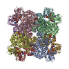
EMDB-29915:
CryoEM map of a de novo designed octahedral nanocage with programmable volume; design cage_O4_34
EMDB-40070:
Cryo-EM map of synthetic cage_O3_10 reconstructed without symmetry (C1)
EMDB-40071:
Cryo-EM map of synthetic cage_O3_10 reconstructed with O symmetry
EMDB-40073:
Cryo-EM map of synthetic cage_T3_5 reconstructed without symmetry (C1), with 1 monomer missing (class 3.0)
EMDB-40074:
Cryo-EM map of synthetic cage_T3_5 reconstructed with T symmetry
EMDB-40075:
Cryo-EM map of synthetic cage_T3_5 reconstructed without symmetry (C1)

EMDB-40076:
Cryo-EM map of synthetic cage_T3_5+2 reconstructed without symmetry (C1)
EMDB-40072:
Cryo-EM map of synthetic cage_T3_5 reconstructed without symmetry (C1), with 1 trimer missing (class 3.1)

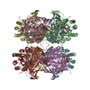
EMDB-26574:
KS-AT di-domain of mycobacterial Pks13 with endogenous KS ligand bound
EMDB-27002:
ACP1-KS-AT domains of mycobacterial Pks13
EMDB-27003:
KS-AT domains of mycobacterial Pks13 with inward AT conformation

EMDB-27004:
KS-AT domains of mycobacterial Pks13 with outward AT conformation
EMDB-27005:
ACP1-KS-AT domains of mycobacterial Pks13

PDB-7uk4:
KS-AT di-domain of mycobacterial Pks13 with endogenous KS ligand bound

PDB-8cuy:
ACP1-KS-AT domains of mycobacterial Pks13

PDB-8cuz:
KS-AT domains of mycobacterial Pks13 with inward AT conformation

PDB-8cv0:
KS-AT domains of mycobacterial Pks13 with outward AT conformation

PDB-8cv1:
ACP1-KS-AT domains of mycobacterial Pks13

EMDB-25825:
AtTPC1 D454N-EDTA state II

EMDB-25826:
AtTPC1 DDE mutant with 1 mM Ca2+

PDB-7tdd:
AtTPC1 D454N-EDTA state II

PDB-7tde:
AtTPC1 DDE mutant with 1 mM Ca2+
EMDB-26839:
KSQ+AT from first module of the pikromycin synthase
EMDB-27094:
AT from first module of the pikromycin synthase

PDB-7uwr:
KSQ+AT from first module of the pikromycin synthase

PDB-8czc:
AT from first module of the pikromycin synthase

EMDB-25798:
AtTPC1 D454N with 1 mM Ca2+

EMDB-25827:
AtTPC1 D454N with 1 mM EDTA state I

PDB-7tbg:
AtTPC1 D454N with 1 mM Ca2+
Pages:
 wwPDB to switch to version 3 of the EMDB data model
