 Movie
Movie Controller

[English] 日本語
 Yorodumi
- EMDB-40071: Cryo-EM map of synthetic cage_O3_10 reconstructed with O symmetry -
+ Open data

- Basic information
| Entry | 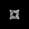 | |||||||||
|---|---|---|---|---|---|---|---|---|---|---|
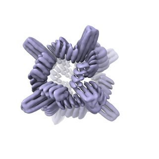
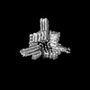
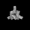
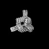
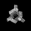
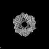
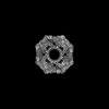
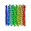
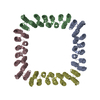
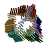
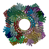
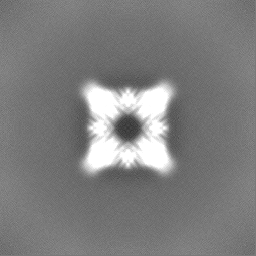
| Title | Cryo-EM map of synthetic cage_O3_10 reconstructed with O symmetry | |||||||||
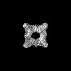
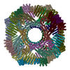
 Map data Map data | cage_O3_10 with O symmetry | |||||||||
 Sample Sample |
| |||||||||
 Keywords Keywords | synthetic / self-assembling / DE NOVO PROTEIN | |||||||||
| Biological species | synthetic construct (others) | |||||||||
| Method | single particle reconstruction / cryo EM / Resolution: 6.0 Å | |||||||||
 Authors Authors | Coudray N / Redler R / Hsia Y / Huddy TF / Baker D / Ekiert D / Bhabha G | |||||||||
| Funding support |  United States, 1 items United States, 1 items
| |||||||||
 Citation Citation | Journal: bioRxiv / Year: 2023 Title: Blueprinting expandable nanomaterials with standardized protein building blocks. Abstract: A wooden house frame consists of many different lumber pieces, but because of the regularity of these building blocks, the structure can be designed using straightforward geometrical principles. The ...A wooden house frame consists of many different lumber pieces, but because of the regularity of these building blocks, the structure can be designed using straightforward geometrical principles. The design of multicomponent protein assemblies in comparison has been much more complex, largely due to the irregular shapes of protein structures . Here we describe extendable linear, curved, and angled protein building blocks, as well as inter-block interactions that conform to specified geometric standards; assemblies designed using these blocks inherit their extendability and regular interaction surfaces, enabling them to be expanded or contracted by varying the number of modules, and reinforced with secondary struts. Using X-ray crystallography and electron microscopy, we validate nanomaterial designs ranging from simple polygonal and circular oligomers that can be concentrically nested, up to large polyhedral nanocages and unbounded straight "train track" assemblies with reconfigurable sizes and geometries that can be readily blueprinted. Because of the complexity of protein structures and sequence-structure relationships, it has not been previously possible to build up large protein assemblies by deliberate placement of protein backbones onto a blank 3D canvas; the simplicity and geometric regularity of our design platform now enables construction of protein nanomaterials according to "back of an envelope" architectural blueprints. | |||||||||
| History |
|
- Structure visualization
| Supplemental images |
|---|
- Downloads & links
-EMDB archive
| Map data | emd_40071.map.gz | 243.9 MB |  EMDB map data format EMDB map data format | |
|---|---|---|---|---|
| Header (meta data) | emd-40071-v30.xmlemd-40071.xml | 23.1 KB 23.1 KB | Display Display | EMDB header |
| Images |  emd_40071.png emd_40071.png | 57.8 KB | ||
| Masks | emd_40071_msk_1.map | 512 MB | Mask map | |
| Filedesc metadata | emd-40071.cif.gz | 6.1 KB | ||
| Others | emd_40071_half_map_1.map.gzemd_40071_half_map_2.map.gz | 475.2 MB 475.2 MB | ||
| Archive directory |  http://ftp.pdbj.org/pub/emdb/structures/EMD-40071ftp://ftp.pdbj.org/pub/emdb/structures/EMD-40071 http://ftp.pdbj.org/pub/emdb/structures/EMD-40071ftp://ftp.pdbj.org/pub/emdb/structures/EMD-40071 | HTTPS FTP |
-Related structure data


-Links
| EMDB pages | EMDB (EBI/PDBe) / EMDataResource |
|---|
-Map
| File | Download / File: emd_40071.map.gz / Format: CCP4 / Size: 512 MB / Type: IMAGE STORED AS FLOATING POINT NUMBER (4 BYTES) | ||||||||||||||||||||||||||||||||||||
|---|---|---|---|---|---|---|---|---|---|---|---|---|---|---|---|---|---|---|---|---|---|---|---|---|---|---|---|---|---|---|---|---|---|---|---|---|---|
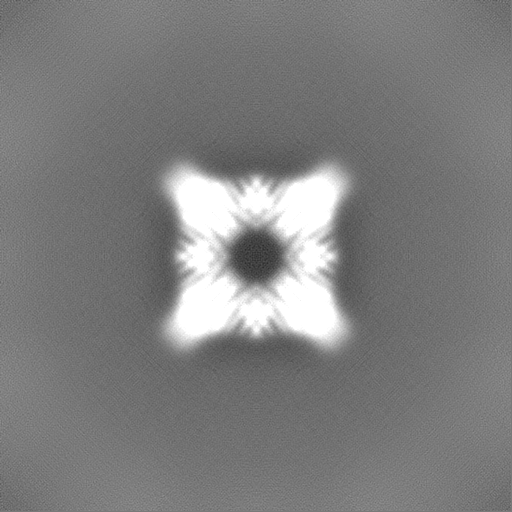
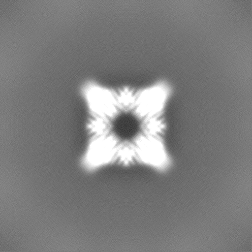
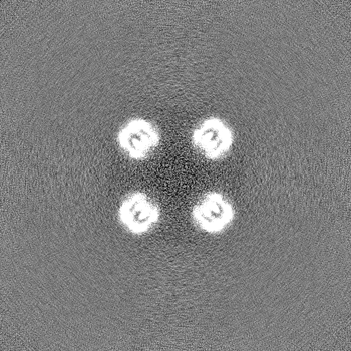
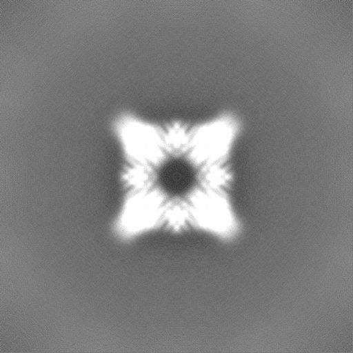
| Annotation | cage_O3_10 with O symmetry | ||||||||||||||||||||||||||||||||||||
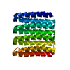
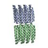
| Projections & slices | Image control
Images are generated by Spider. | ||||||||||||||||||||||||||||||||||||
| Voxel size | X=Y=Z: 0.8248 Å | ||||||||||||||||||||||||||||||||||||
| Density |
| ||||||||||||||||||||||||||||||||||||
| Symmetry | Space group: 1 | ||||||||||||||||||||||||||||||||||||
| Details | EMDB XML:
|
 Z (Sec.)
 Y (Row.)
 X (Col.)


-Supplemental data
-Mask #1
| File | emd_40071_msk_1.map | ||||||||||||
|---|---|---|---|---|---|---|---|---|---|---|---|---|---|
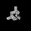
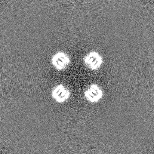
| Projections & Slices |
| ||||||||||||
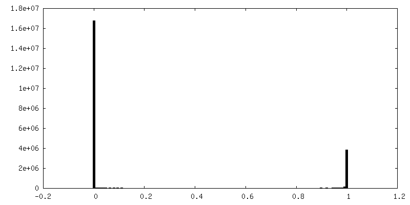
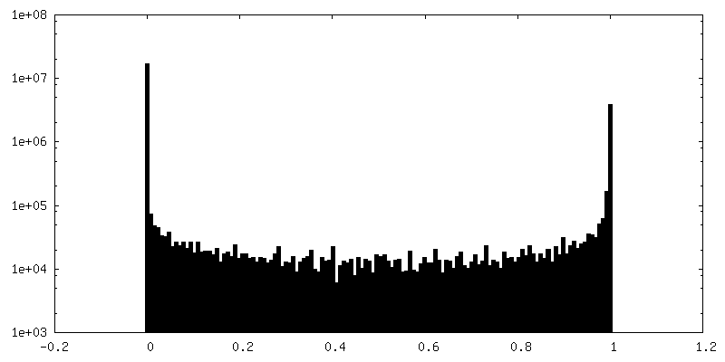
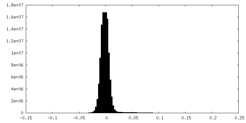
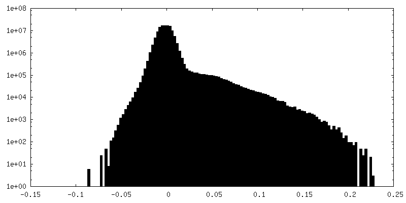
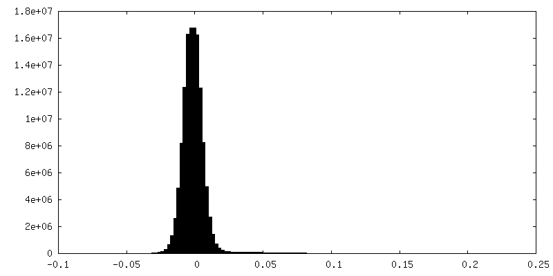
| Density Histograms |


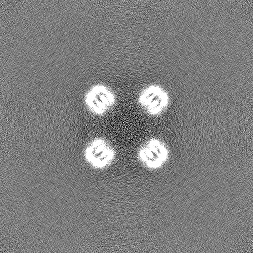
-Half map: cage O3 10 with O symmetry, half map A
| File | emd_40071_half_map_1.map | ||||||||||||
|---|---|---|---|---|---|---|---|---|---|---|---|---|---|
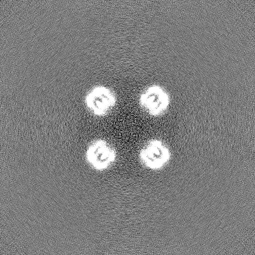
| Annotation | cage_O3_10 with O symmetry, half map A | ||||||||||||
| Projections & Slices |
| ||||||||||||
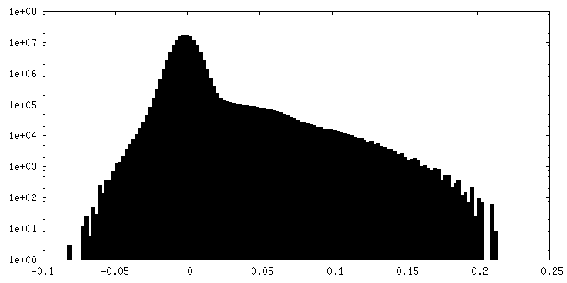
| Density Histograms |

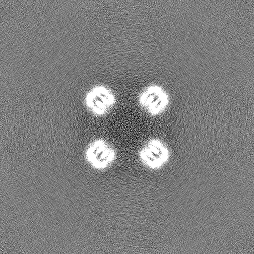
-Half map: cage O3 10 with O symmetry, half map B
| File | emd_40071_half_map_2.map | ||||||||||||
|---|---|---|---|---|---|---|---|---|---|---|---|---|---|
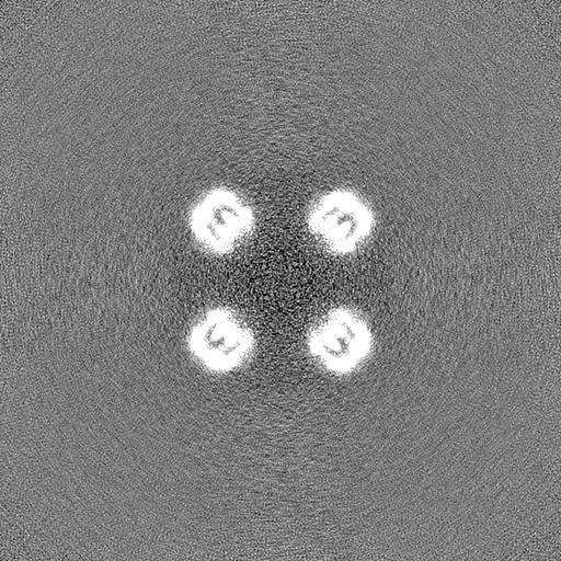
| Annotation | cage_O3_10 with O symmetry, half map B | ||||||||||||
| Projections & Slices |
| ||||||||||||
| Density Histograms |

- Sample components
-Entire : Expandable de novo designed complex cage_O3_10
| Entire | Name: Expandable de novo designed complex cage_O3_10 |
|---|---|
| Components |
|
-Supramolecule #1: Expandable de novo designed complex cage_O3_10
| Supramolecule | Name: Expandable de novo designed complex cage_O3_10 / type: complex / ID: 1 / Parent: 0 / Macromolecule list: all |
|---|---|
| Source (natural) | Organism: synthetic construct (others) |
-Macromolecule #1: cage_03_10
| Macromolecule | Name: cage_03_10 / type: protein_or_peptide / ID: 1 / Details: The cage contains 24 copies of the monomer. / Enantiomer: LEVO |
|---|---|
| Source (natural) | Organism: synthetic construct (others) |
| Recombinant expression | Organism:  |
| Sequence | String: mGSVELLAVA ALQELNIELA RALLEAVARL QELNIDLVRK TSELTDEKTI REEIRKVKEE SKRIVEEAEE LIRLAKLASE AIARMAEVAA RGAPPELLIE LLERLLKKAQ EAGMSPEIIH LLLELALAIV EARGVPPEQL AEFAERLVEI LREAGGSPEL VFELLQRIMK ...String: mGSVELLAVA ALQELNIELA RALLEAVARL QELNIDLVRK TSELTDEKTI REEIRKVKEE SKRIVEEAEE LIRLAKLASE AIARMAEVAA RGAPPELLIE LLERLLKKAQ EAGMSPEIIH LLLELALAIV EARGVPPEQL AEFAERLVEI LREAGGSPEL VFELLQRIMK IIERRGAPPE LLIELLERLL ELAREAGLSP EQILRLLITA LLIVSKRGVP PEQLAEFAEK LKEILREAGG SPEAQEALKR LIEAIEDLRG AGGSGGSGGS lehhhhhh |
-Experimental details
-Structure determination
| Method | cryo EM |
|---|---|
 Processing Processing | single particle reconstruction |
| Aggregation state | particle |
-Sample preparation
| Concentration | 0.5 mg/mL |
|---|---|
| Buffer | pH: 8 |
| Grid | Model: Quantifoil R2/2 / Material: COPPER / Mesh: 300 / Support film - Material: CARBON / Support film - topology: CONTINUOUS / Support film - Film thickness: 2 / Pretreatment - Type: GLOW DISCHARGE / Pretreatment - Time: 5 sec. |
| Vitrification | Cryogen name: ETHANE / Chamber humidity: 100 % / Chamber temperature: 295 K / Instrument: FEI VITROBOT MARK IV |
- Electron microscopy
| Microscope | FEI TITAN KRIOS |
|---|---|
| Specialist optics | Energy filter - Slit width: 30 eV |
| Image recording | Film or detector model: GATAN K3 (6k x 4k) / Digitization - Dimensions - Width: 5760 pixel / Digitization - Dimensions - Height: 4092 pixel / Number grids imaged: 2 / Number real images: 4262 / Average exposure time: 2.0 sec. / Average electron dose: 58.8 e/Å2 |
| Electron beam | Acceleration voltage: 300 kV / Electron source:  FIELD EMISSION GUN FIELD EMISSION GUN |
| Electron optics | Illumination mode: FLOOD BEAM / Imaging mode: BRIGHT FIELD / Cs: 2.7 mm / Nominal defocus max: 2.2 µm / Nominal defocus min: 0.6 µm / Nominal magnification: 105000 |
| Sample stage | Specimen holder model: FEI TITAN KRIOS AUTOGRID HOLDER |
| Experimental equipment |  Model: Titan Krios / Image courtesy: FEI Company |
+Image processing
-Atomic model buiding 1
| Initial model | Chain - Source name: Other / Chain - Initial model type: in silico model / Details: de novo designed model |
|---|
