 Movie
Movie Controller
 Structure viewers
 About EMN search


-Search query
-Search result
Showing 1 - 50 of 459 items for (author: dick & m)
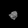
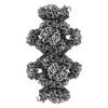
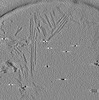
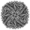
EMDB-50504: 
in situ 70S Myxococcus xanthus ribosome
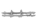
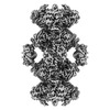
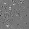
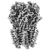
EMDB-50515:
PomX filament from Myxococcus xanthus
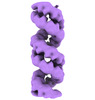
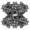
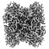
EMDB-19599:
Structural characterization of Thogoto Virus nucleoprotein provides insights into RNA encapsidation and assembly
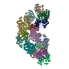
PDB-8ryt:
Structural characterization of Thogoto Virus nucleoprotein provides insights into RNA encapsidation and assembly

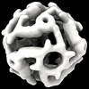
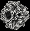
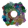
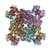
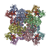
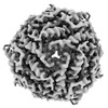
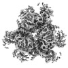
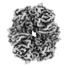
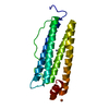
EMDB-41907:
Computationally Designed, Expandable O4 Octahedral Handshake Nanocage
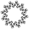
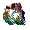
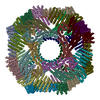
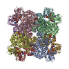
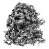
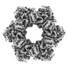
EMDB-42031:
Computational Designed Nanocage O43_129_+8
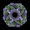
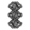
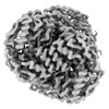
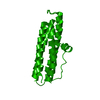
EMDB-43318:
Twistless helix 12 repeat ring design R12B
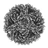
EMDB-29974:
Cryo-EM structure of synthetic tetrameric building block sC4
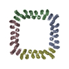
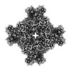
EMDB-41364:
CryoEM Structure of a Computationally Designed T3 Tetrahedral Nanocage
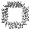
EMDB-42906:
Computational Designed Nanocage O43_129
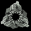
EMDB-42944:
Computational Designed Nanocage O43_129_+4
PDB-8gel:
Cryo-EM structure of synthetic tetrameric building block sC4
PDB-8tl7:
CryoEM Structure of a Computationally Designed T3 Tetrahedral Nanocage
PDB-8v2d:
Computational Designed Nanocage O43_129
PDB-8v3b:
Computational Designed Nanocage O43_129_+4
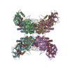
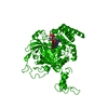
EMDB-41986:
Human retinal variant phosphomimetic IMPDH1(595)-S477D free octamer bound by GTP, ATP, IMP, and NAD+
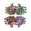
EMDB-41989:
Human retinal variant phosphomimetic IMPDH1(546)-S477D filament bound by GTP, ATP, IMP, and NAD+, octamer-centered
EMDB-42012:
Human retinal variant phosphomimetic IMPDH1(546)-S477D filament bound by GTP, ATP, IMP, and NAD+, interface-centered
EMDB-42026:
Human retinal variant phosphomimetic IMPDH1(546)-S477D filament bound by ATP, IMP, and NAD+, octamer-centered
EMDB-42029:
Human retinal variant phosphomimetic IMPDH1(546)-S477D filament bound by ATP, IMP, and NAD+, interface-centered
PDB-8u7m:
Human retinal variant phosphomimetic IMPDH1(595)-S477D free octamer bound by GTP, ATP, IMP, and NAD+
PDB-8u7q:
Human retinal variant phosphomimetic IMPDH1(546)-S477D filament bound by GTP, ATP, IMP, and NAD+, octamer-centered
PDB-8u7v:
Human retinal variant phosphomimetic IMPDH1(546)-S477D filament bound by GTP, ATP, IMP, and NAD+, interface-centered
PDB-8u8o:
Human retinal variant phosphomimetic IMPDH1(546)-S477D filament bound by ATP, IMP, and NAD+, octamer-centered
PDB-8u8y:
Human retinal variant phosphomimetic IMPDH1(546)-S477D filament bound by ATP, IMP, and NAD+, interface-centered
EMDB-28752:
Cryo-electron tomography of wild type a-synuclein preformed fibrils
EMDB-28753:
Cryo-electron tomography of S42Y a-synuclein preformed fibrils

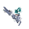
EMDB-18941:
SARS-CoV-2 S (Spike) protein (BA.1) in complex with VHH Ma16B06 (sub-volume of two adjacent RBD-VHH modules)
EMDB-17958:
Structure of DPS determined by cryoEM at 100 keV
EMDB-17959:
Structure of bacterial ribosome determined by cryoEM at 100 keV
EMDB-17960:
Structure of GABAAR determined by cryoEM at 100 keV
EMDB-17961:
Structure of mouse heavy-chain apoferritin determined by cryoEM at 100 keV
EMDB-17962:
Structure of catalase determined by cryoEM at 100 keV
EMDB-17963:
Structure of AHIR determined by cryoEM at 100 keV
EMDB-17964:
Structure of GAPDH determined by cryoEM at 100 keV
EMDB-17965:
Structure of E. coli glutamine synthetase determined by cryoEM at 100 keV
EMDB-17966:
Structure of human apo ALDH1A1 determined by cryoEM at 100 keV

EMDB-17967:
Structure of PaaZ determined by cryoEM at 100 keV
EMDB-17968:
Structure of lumazine synthase determined by cryoEM at 100 keV
PDB-8pv9:
Structure of DPS determined by cryoEM at 100 keV

PDB-8pva:
Structure of bacterial ribosome determined by cryoEM at 100 keV
PDB-8pvb:
Structure of GABAAR determined by cryoEM at 100 keV
PDB-8pvc:
Structure of mouse heavy-chain apoferritin determined by cryoEM at 100 keV
PDB-8pvd:
Structure of catalase determined by cryoEM at 100 keV
PDB-8pve:
Structure of AHIR determined by cryoEM at 100 keV
PDB-8pvf:
Structure of GAPDH determined by cryoEM at 100 keV
PDB-8pvg:
Structure of E. coli glutamine synthetase determined by cryoEM at 100 keV
PDB-8pvh:
Structure of human apo ALDH1A1 determined by cryoEM at 100 keV

PDB-8pvi:
Structure of PaaZ determined by cryoEM at 100 keV
PDB-8pvj:
Structure of lumazine synthase determined by cryoEM at 100 keV
Pages:
 wwPDB to switch to version 3 of the EMDB data model
