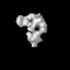
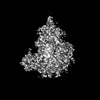
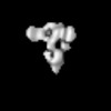
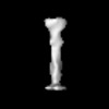
 Movie
Movie Controller
 Structure viewers
 About EMN search


-Search query
-Search result
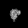
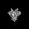
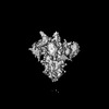
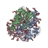
Showing 1 - 50 of 53 items for (author: cheng & zk)

EMDB-63216: 
Cryo-EM structure of prefusion-stabilized RSV F (DS-Cav1 strain: A2) in complex with nanobody 1G9
Method: single particle / : Wang QQ, Ke XL, Li ET, Hong DX, Li HX, Cheng ZK, Zhang JC, Jin TC, Shu B, Chiu S

EMDB-63217:
Cryo-EM structure of prefusion-stabilized RSV F (DS-Cav1 strain: A2) in complex with nanobody 1D8
Method: single particle / : Wang QQ, Ke XL, Li ET, Hong DX, Li HX, Cheng ZK, Zhang JC, Jin TC, Shu B, Chiu S
EMDB-40786:
Structural basis and functional roles for Toll-like receptor binding to Latrophilin adhesion-GPCR in embryo development
Method: single particle / : Li J, Rosas GC, Arac D, Ozkan E

EMDB-27640:
Org 2274179-0-bound Thyrotropin Receptor
Method: single particle / : Faust B, Cheng Y, Manglik A

EMDB-27647:
Human thyrotropin analog TR1402 bound to human Thyrotropin receptor (extracellular domain map only)
Method: single particle / : Faust B, Cheng Y, Manglik A

EMDB-27648:
Human thyrotropin analog TR1402 bound to human Thyrotropin receptor (transmembrane domain and G protein map only)
Method: single particle / : Faust B, Cheng Y, Manglik A

EMDB-27649:
Native human TSH bound to human Thyrotropin receptor (extracellular domain map only)
Method: single particle / : Faust B, Cheng Y, Manglik A

EMDB-27650:
Native human TSH bound to human Thyrotropin receptor (transmembrane domain and G protein map only)
Method: single particle / : Faust B, Cheng Y, Manglik A

EMDB-27651:
M22 Fab bound to human Thyrotropin receptor (extracellular domain map only)
Method: single particle / : Faust B, Cheng Y, Manglik A

EMDB-27652:
M22 Fab bound to human Thyrotropin receptor (transmembrane domain and G protein map only)
Method: single particle / : Faust B, Cheng Y, Manglik A

EMDB-25762:
Human Thyrotropin receptor bound by CS-17 Inverse Agonist Fab/Org 274179-0 Antagonist
Method: single particle / : Faust B, Cheng Y, Manglik A

PDB-7t9m:
Human Thyrotropin receptor bound by CS-17 Inverse Agonist Fab/Org 274179-0 Antagonist
Method: single particle / : Faust B, Cheng Y, Manglik A
EMDB-32329:
Cryo-EM map of PEDV (Pintung 52) S protein with all three protomers in the D0-down conformation determined in situ on intact viral particles.
Method: single particle / : Hsu STD, Draczkowski P
EMDB-32332:
Subtomogram averaging of PEDV (Pintung 52) S protein with all three protomers in the D0-down conformation determined in situ on intact viral particles.
Method: subtomogram averaging / : Hsu STD, Draczkowski P, Wang YS, Huang CY
EMDB-32333:
Subtomogram averaging of PEDV (Pintung 52) S protein with one protomer in the D0-up conformation and two protomers in the D0-down conformation, determined in situ on intact viral particles
Method: subtomogram averaging / : Hsu STD, Draczkowski P, Wang YS, Huang CY
EMDB-32337:
Subtomogram averaging of PEDV (Pintung 52) S protein with two protomers in the D0-up conformation and one protomer in the D0-down conformation, determined in situ on intact viral particles.
Method: subtomogram averaging / : Hsu STD, Draczkowski P, Wang YS, Huang CY
EMDB-32338:
Cryo-EM map of PEDV S protein with one protomer in the D0-up conformation while the other two in the D0-down conformation
Method: single particle / : Hsu STD, Draczkowski P, Wang YS
EMDB-32339:
Subtomogram averaging of PEDV (Pintung 52) S protein with all three protomers in the D0-up conformation determined in situ on intact viral particles.
Method: subtomogram averaging / : Hsu STD, Draczkowski P, Wang YS, Huang CY
EMDB-32340:
Subtomogram averaging of PEDV (Pintung 52) S protein in the postfusion form determined in situ on intact viral particles.
Method: subtomogram averaging / : Hsu STD, Draczkowski P, Wang YS, Huang CY
EMDB-33646:
Cryo-EM map of IPEC-J2 cell-derived PEDV PT52 S protein with three D0-up
Method: single particle / : Hsu STD, Draczkowski P, Wang YS
EMDB-33647:
Cryo-EM map of IPEC-J2 cell-derived PEDV PT52 S protein one D0-down and two D0-up
Method: single particle / : Hsu STD, Draczkowski P, Wang YS
EMDB-33648:
Symmetry-expanded and locally refined protomer structure of IPEC-J2 cell-derived PEDV PT52 S with a CTD-close conformation
Method: single particle / : Hsu STD, Draczkowski P, Wang YS
EMDB-33649:
Symmetry-expanded and locally refined protomer structure of IPEC-J2 cell-derived PEDV PT52 S with a CTD-open conformation
Method: single particle / : Hsu STD, Draczkowski P, Wang YS
EMDB-33700:
Cryo-EM map of HEK293F cell-derived PEDV PT52 S protein with three D0-down
Method: single particle / : Hsu STD, Draczkowski P, Wang YS
EMDB-33701:
Cryo-EM map of HEK293F cell-derived PEDV PT52 S protein one D0-up and two D0-down
Method: single particle / : Hsu STD, Draczkowski P, Wang YS
EMDB-33702:
Cryo-EM map of HEK293F cell-derived PEDV PT52 S protein with three D0-up
Method: single particle / : Hsu STD, Draczkowski P, Wang YS
EMDB-33703:
Cryo-EM map of HEK293F cell-derived PEDV PT52 S T326I with three D0-down
Method: single particle / : Hsu STD, Draczkowski P, Wang YS
EMDB-33704:
Cryo-EM map of HEK293F cell-derived PEDV PT52 S T326I one D0-up and two D0-down
Method: single particle / : Hsu STD, Draczkowski P, Wang YS
EMDB-33705:
Cryo-EM map of HEK293F cell-derived PEDV PT52 S T326I one D0-down and two D0-up
Method: single particle / : Hsu STD, Draczkowski P, Wang YS
EMDB-33706:
Cryo-EM map of HEK293F cell-derived PEDV PT52 S T326I with three D0-up
Method: single particle / : Hsu STD, Draczkowski P, Wang YS
PDB-7w6m:
Cryo-EM map of PEDV (Pintung 52) S protein with all three protomers in the D0-down conformation determined in situ on intact viral particles.
Method: single particle / : Hsu STD, Draczkowski P, Wang YS
PDB-7w73:
Cryo-EM map of PEDV S protein with one protomer in the D0-up conformation while the other two in the D0-down conformation
Method: single particle / : Hsu STD, Draczkowski P, Wang YS

PDB-7y6s:
Cryo-EM map of IPEC-J2 cell-derived PEDV PT52 S protein with three D0-up
Method: single particle / : Hsu STD, Draczkowski P, Wang YS
PDB-7y6t:
Cryo-EM map of IPEC-J2 cell-derived PEDV PT52 S protein one D0-down and two D0-up
Method: single particle / : Hsu STD, Draczkowski P, Wang YS
PDB-7y6u:
Symmetry-expanded and locally refined protomer structure of IPEC-J2 cell-derived PEDV PT52 S with a CTD-close conformation
Method: single particle / : Hsu STD, Draczkowski P, Wang YS
PDB-7y6v:
Symmetry-expanded and locally refined protomer structure of IPEC-J2 cell-derived PEDV PT52 S with a CTD-open conformation
Method: single particle / : Hsu STD, Draczkowski P, Wang YS
EMDB-25758:
Native human TSH bound to human Thyrotropin receptor in complex with miniGs399 (composite structure)
Method: single particle / : Faust B, Cheng Y, Manglik A

EMDB-25763:
M22 Agonist Autoantibody bound to Human Thyrotropin receptor in complex with miniGs399 (composite structure)
Method: single particle / : Faust B, Cheng Y, Manglik A
EMDB-26795:
Human thyrotropin analog TR1402 bound to human Thyrotropin receptor in complex with miniGs399 (composite structure)
Method: single particle / : Faust B, Cheng Y, Manglik A
PDB-7t9i:
Native human TSH bound to human Thyrotropin receptor in complex with miniGs399 (composite structure)
Method: single particle / : Faust B, Cheng Y, Manglik A
PDB-7t9n:
M22 Agonist Autoantibody bound to Human Thyrotropin receptor in complex with miniGs399 (composite structure)
Method: single particle / : Faust B, Cheng Y, Manglik A

PDB-7utz:
Human thyrotropin analog TR1402 bound to human Thyrotropin receptor in complex with miniGs399 (composite structure)
Method: single particle / : Faust B, Cheng Y, Manglik A
EMDB-7541:
Rabbit muscle aldolase at 2.4A resolution (17dec27a 205k particles, all images)
Method: single particle / : Kim LK

EMDB-7550:
Rabbit muscle aldolase at 2.4A resolution (17dec27a 205k particles, all images)
Method: single particle / : Kim LK, Rice WJ, Eng ET, Kopylov M, Cheng A, Raczkowski AM, Jordan KD, Bobe D, Potter CS, Carragher B
EMDB-7551:
Rabbit muscle aldolase at 3.0A resolution (17nov02c all img, 204k particles)
Method: single particle / : Kim LK, Rice WJ, Eng ET, Kopylov M, Cheng A, Raczkowski AM, Jordan KD, Bobe D, Potter CS, Carragher B

EMDB-7562:
Rabbit muscle aldolase at 3.5 A resolution (17nov02c less 25nm ice thickness, 22k particles)
Method: single particle / : Kim LK, Rice WJ, Eng ET, Kopylov M, Cheng A, Raczkowski AM, Jordan KD, Bobe D, Potter CS, Carragher B
EMDB-7614:
Rabbit muscle aldolase at 2.8 A resolution (17sep21j 1st 500 img, 62k particles)
Method: single particle / : Kim LK, Rice WJ, Eng ET, Kopylov M, Cheng A, Raczkowski AM, Jordan KD, Bobe D, Potter CS, Carragher B
EMDB-7615:
Rabbit muscle aldolase at 4.6 A resolution (17nov02c 1st700 img, 75k particles)
Method: single particle / : Kim LK, Rice WJ, Eng ET, Kopylov M, Cheng A, Raczkowski AM, Jordan KD, Bobe D, Potter CS, Carragher B

EMDB-7616:
Rabbit muscle aldolase at 2.5 A resolution (17sep21j all img, 219k particles)
Method: single particle / : Kim LK, Rice WJ, Eng ET, Kopylov M, Cheng A, Raczkowski AM, Jordan KD, Bobe D, Potter CS, Carragher B
EMDB-7617:
Rabbit muscle aldolase at 2.5 A resolution (17sep21j less 25nm ice thickness, 124k particles)
Method: single particle / : Kim LK, Rice WJ, Eng ET, Kopylov M, Cheng A, Raczkowski AM, Jordan KD, Bobe D, Potter CS, Carragher B
Pages:
 wwPDB to switch to version 3 of the EMDB data model
