 ムービー
ムービー コントローラー
 構造ビューア
 EMN検索について


-検索条件
-検索結果
検索 (著者・登録者: bruno & ec)の結果全46件を表示しています

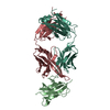
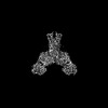
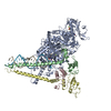
EMDB-65225: 
Cryo-EM Structure of Human GPR158 Bound to Nanobody Nb20
PDB-9vor:
Cryo-EM Structure of Human GPR158 Bound to Nanobody Nb20

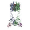
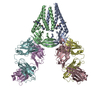
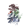
EMDB-61843:
Yeast Mitochondrial PORIN complex

PDB-9jvq:
Yeast Mitochondrial PORIN complex

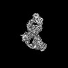
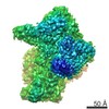
EMDB-53511:
SpCas9 with computationally designed SpCas9_b10 binder
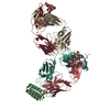
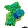
EMDB-53510:
SpCas9 with computationally designed SpCas9_b3 binder
EMDB-52864:
CryoEM structure of a synthetic antibody, COP-2, in complex with the C-terminal domain of Clostridium perfringens Enterotoxin
PDB-9ihc:
CryoEM structure of a synthetic antibody, COP-2, in complex with the C-terminal domain of Clostridium perfringens Enterotoxin

EMDB-50034:
SARS-CoV-2 M protein dimer (short form) in complex with Fab-B and CIM-834
EMDB-50035:
SARS-CoV-2 M protein dimer (long form) in complex with Fab-E and incubated with CIM-834
PDB-9exa:
SARS-CoV-2 M protein dimer (short form) in complex with Fab-B and CIM-834
EMDB-50522:
Progesterone-bound DB3 Fab in complex with computationally designed DBPro1156_2 protein binder
PDB-9fkd:
Progesterone-bound DB3 Fab in complex with computationally designed DBPro1156_2 protein binder
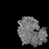
EMDB-18539:
Structure of the human 80S ribosome at 1.9 A resolution - the molecular role of chemical modifications and ions in RNA
EMDB-18812:
The structure of the human 80S ribosome at 1.9 angstrom resolution reveals the molecular role of chemical modifications and ions in RNA - Focused refinement of the of the 60S subunit
EMDB-18813:
The structure of the human 80S ribosome at 1.9 angstrom resolution reveals the molecular role of chemical modifications and ions in RNA - Focused refinement of the of the 40S subunit body
EMDB-18814:
The structure of the human 80S ribosome at 1.9 angstrom resolution reveals the molecular role of chemical modifications and ions in RNA - Focused refinement of the of the 40S subunit head
EMDB-18815:
Structure of the human 80S ribosome at 1.9 A resolution - the molecular role of chemical modifications and ions in RNA - Global human 80S ribosome refinement before focused refinements.
PDB-8qoi:
Structure of the human 80S ribosome at 1.9 A resolution - the molecular role of chemical modifications and ions in RNA

EMDB-44479:
Cryo-EM structure of synthetic claudin-4 complex with Clostridium perfringens enterotoxin C-terminal domain, sFab COP-2, and Nanobody

PDB-9bei:
Cryo-EM structure of synthetic claudin-4 complex with Clostridium perfringens enterotoxin C-terminal domain, sFab COP-2, and Nanobody
EMDB-14922:
cryo-EM structure of omicron spike in complex with de novo designed binder, full map
PDB-7zrv:
cryo-EM structure of omicron spike in complex with de novo designed binder, full map
EMDB-14930:
cryo-EM structure of omicron spike in complex with de novo designed binder, local

EMDB-14947:
cryo-EM structure of D614 spike in complex with de novo designed binder, full and local maps(addition)
PDB-7zsd:
cryo-EM structure of omicron spike in complex with de novo designed binder, local
PDB-7zss:
cryo-EM structure of D614 spike in complex with de novo designed binder
EMDB-31061:
A dual mechanism of action of AT-527 against SARS-CoV-2 polymerase
PDB-7ed5:
A dual mechanism of action of AT-527 against SARS-CoV-2 polymerase

EMDB-12798:
Hexameric coxsackievirus B3 2C protein in complex with S-fluoxetine

EMDB-12158:
CryoEM structure of Mycobacterium tuberculosis UMP Kinase (UMPK) in complex with UDP and UTP
PDB-7bes:
CryoEM structure of Mycobacterium tuberculosis UMP Kinase (UMPK) in complex with UDP and UTP
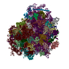
EMDB-8149:
Cryo-EM structure of a full archaeal ribosomal translation initiation complex in the P-INconformation

PDB-5jbh:
Cryo-EM structure of a full archaeal ribosomal translation initiation complex in the P-IN conformation
EMDB-8148:
Cryo-EM structure of a full archaeal ribosomal translation initiation complex in the P-REMOTE conformation

PDB-5jb3:
Cryo-EM structure of a full archaeal ribosomal translation initiation complex in the P-REMOTE conformation
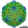
EMDB-2816:
Electron cryoEM structure of lactococcal siphophage 1358 virion
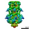
EMDB-2817:
Electron cryoEM structure of lactococcal siphophage 1358 virion
EMDB-2819:
Electron cryoEM structure of lactococcal siphophage 1358 virion
EMDB-2820:
Electron cryoEM structure of lactococcal siphophage 1358 virion
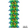
EMDB-2698:
The cryoEM structure of Monalysin Toxin
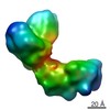
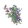
EMDB-2631:
The Cryo-EM structure of the palindromic DNA-bound USP/EcR nuclear receptor reveals an asymmetric organization with allosteric domain positioning
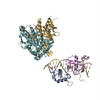
PDB-4umm:
The Cryo-EM structure of the palindromic DNA-bound USP-EcR nuclear receptor reveals an asymmetric organization with allosteric domain positioning
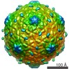
EMDB-2647:
electron cryo-microscopy of 1358 Lactococcus phage mature empty capsid
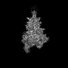
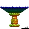
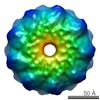
EMDB-1188:
An archaeal peptidase assembles into two different quaternary structures: A tetrahedron and a giant octahedron.
EMDB-1189:
An archaeal peptidase assembles into two different quaternary structures: A tetrahedron and a giant octahedron.
 wwPDBはEMDBデータモデルのバージョン3へ移行します
