 Movie
Movie Controller

[English] 日本語
 Yorodumi
- PDB-9fkd: Progesterone-bound DB3 Fab in complex with computationally design... -
+ Open data

- Basic information
| Entry | Database: PDB / ID: 9fkd | ||||||
|---|---|---|---|---|---|---|---|
| Title | Progesterone-bound DB3 Fab in complex with computationally designed DBPro1156_2 protein binder | ||||||
 Components Components |
| ||||||
 Keywords Keywords | DE NOVO PROTEIN / progesterone / binder / de novo / Fab / anti-kappa | ||||||
| Function / homology | PROGESTERONE Function and homology information Function and homology information | ||||||
| Biological species | synthetic construct (others) | ||||||
| Method | ELECTRON MICROSCOPY / single particle reconstruction / cryo EM / Resolution: 3.3 Å | ||||||
 Authors Authors | Pacesa, M. / Marchand, A. / Correia, B.E. | ||||||
| Funding support |  Switzerland, 1items Switzerland, 1items
| ||||||
 Citation Citation | Journal: Nature / Year: 2025 Title: Targeting protein-ligand neosurfaces with a generalizable deep learning tool. Authors: Anthony Marchand / Stephen Buckley / Arne Schneuing / Martin Pacesa / Maddalena Elia / Pablo Gainza / Evgenia Elizarova / Rebecca M Neeser / Pao-Wan Lee / Luc Reymond / Yangyang Miao / Leo ...Authors: Anthony Marchand / Stephen Buckley / Arne Schneuing / Martin Pacesa / Maddalena Elia / Pablo Gainza / Evgenia Elizarova / Rebecca M Neeser / Pao-Wan Lee / Luc Reymond / Yangyang Miao / Leo Scheller / Sandrine Georgeon / Joseph Schmidt / Philippe Schwaller / Sebastian J Maerkl / Michael Bronstein / Bruno E Correia /    Abstract: Molecular recognition events between proteins drive biological processes in living systems. However, higher levels of mechanistic regulation have emerged, in which protein-protein interactions are ...Molecular recognition events between proteins drive biological processes in living systems. However, higher levels of mechanistic regulation have emerged, in which protein-protein interactions are conditioned to small molecules. Despite recent advances, computational tools for the design of new chemically induced protein interactions have remained a challenging task for the field. Here we present a computational strategy for the design of proteins that target neosurfaces, that is, surfaces arising from protein-ligand complexes. To develop this strategy, we leveraged a geometric deep learning approach based on learned molecular surface representations and experimentally validated binders against three drug-bound protein complexes: Bcl2-venetoclax, DB3-progesterone and PDF1-actinonin. All binders demonstrated high affinities and accurate specificities, as assessed by mutational and structural characterization. Remarkably, surface fingerprints previously trained only on proteins could be applied to neosurfaces induced by interactions with small molecules, providing a powerful demonstration of generalizability that is uncommon in other deep learning approaches. We anticipate that such designed chemically induced protein interactions will have the potential to expand the sensing repertoire and the assembly of new synthetic pathways in engineered cells for innovative drug-controlled cell-based therapies. | ||||||
| History |
|
- Structure visualization
| Structure viewer | Molecule: MolmilJmol/JSmol |
|---|
- Downloads & links
-Download
| PDBx/mmCIF format | 9fkd.cif.gz | 206.5 KB | Display | PDBx/mmCIF format |
|---|---|---|---|---|
| PDB format | pdb9fkd.ent.gz | 148.1 KB | Display | PDB format |
| PDBx/mmJSON format | 9fkd.json.gz | Tree view | PDBx/mmJSON format | |
| Others |  Other downloads Other downloads |
-Validation report
| Arichive directory | https://data.pdbj.org/pub/pdb/validation_reports/fk/9fkdftp://data.pdbj.org/pub/pdb/validation_reports/fk/9fkd | HTTPS FTP |
|---|
-Related structure data
| Related structure data | 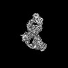 50522MC  8s1xC M: map data used to model this data C: citing same article ( |
|---|---|
| Similar structure data |
-Links
 PDBj
PDBj


- Assembly
| Deposited unit | 
|
|---|---|
| 1 |
|
-Components
-Antibody , 4 types, 4 molecules HLKI
| #2: Antibody | Mass: 25930.930 Da / Num. of mol.: 1 Source method: isolated from a genetically manipulated source Source: (gene. exp.) synthetic construct (others) / Production host:  Homo sapiens (human) Homo sapiens (human) |
|---|---|
| #3: Antibody | Mass: 24332.191 Da / Num. of mol.: 1 Source method: isolated from a genetically manipulated source Source: (gene. exp.) synthetic construct (others) / Production host: Homo sapiens (human) |
| #4: Antibody | Mass: 24115.680 Da / Num. of mol.: 1 Source method: isolated from a genetically manipulated source Source: (gene. exp.) synthetic construct (others) / Production host: Homo sapiens (human) |
| #5: Antibody | Mass: 25346.602 Da / Num. of mol.: 1 Source method: isolated from a genetically manipulated source Source: (gene. exp.) synthetic construct (others) / Production host: Homo sapiens (human) |
-Protein / Non-polymers , 2 types, 2 molecules B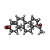
| #1: Protein | Mass: 8087.193 Da / Num. of mol.: 1 Source method: isolated from a genetically manipulated source Source: (gene. exp.) synthetic construct (others) / Production host:  |
|---|---|
| #6: Chemical | ChemComp-STR /  Mass: 314.462 Da / Num. of mol.: 1 / Source method: obtained synthetically / Formula: C21H30O2 / Feature type: SUBJECT OF INVESTIGATION Mass: 314.462 Da / Num. of mol.: 1 / Source method: obtained synthetically / Formula: C21H30O2 / Feature type: SUBJECT OF INVESTIGATION |
-Details
| Has ligand of interest | Y |
|---|---|
| Has protein modification | Y |
-Experimental details
-Experiment
| Experiment | Method: ELECTRON MICROSCOPY |
|---|---|
| EM experiment | Aggregation state: PARTICLE / 3D reconstruction method: single particle reconstruction |
- Sample preparation
| Component | Name: Progesterone-bound DB3 Fab in complex with computationally designed DBPro1156_2 protein binder Type: COMPLEX / Entity ID: #1-#5 / Source: RECOMBINANT |
|---|---|
| Molecular weight | Value: 0.10761566 MDa / Experimental value: NO |
| Source (natural) | Organism: synthetic construct (others) |
| Source (recombinant) | Organism: Homo sapiens (human) |
| Buffer solution | pH: 7.5 |
| Specimen | Conc.: 3.9 mg/ml / Embedding applied: NO / Shadowing applied: NO / Staining applied: NO / Vitrification applied: YES Details: DB3 Fab and anti-Kappa Fab were pre-purified on SEC, while DBPro1156_2 de novo binder was added in 3-fold excess before grid freezing |
| Specimen support | Grid material: GOLD / Grid mesh size: 300 divisions/in. / Grid type: Quantifoil |
| Vitrification | Instrument: FEI VITROBOT MARK IV / Cryogen name: ETHANE / Humidity: 95 % / Chamber temperature: 283.15 K |
- Electron microscopy imaging
| Experimental equipment |  Model: Titan Krios / Image courtesy: FEI Company |
|---|---|
| Microscopy | Model: TFS KRIOS |
| Electron gun | Electron source:  FIELD EMISSION GUN / Accelerating voltage: 300 kV / Illumination mode: FLOOD BEAM FIELD EMISSION GUN / Accelerating voltage: 300 kV / Illumination mode: FLOOD BEAM |
| Electron lens | Mode: BRIGHT FIELD / Nominal defocus max: 2400 nm / Nominal defocus min: 1200 nm / Cs: 2.7 mm / C2 aperture diameter: 50 µm / Alignment procedure: COMA FREE |
| Specimen holder | Cryogen: NITROGEN / Temperature (max): 192 K / Temperature (min): 186 K |
| Image recording | Electron dose: 60 e/Å2 / Film or detector model: FEI FALCON IV (4k x 4k) |
| Image scans | Width: 4096 / Height: 4096 |
- Processing
| EM software |
| ||||||||||||||||||||||||
|---|---|---|---|---|---|---|---|---|---|---|---|---|---|---|---|---|---|---|---|---|---|---|---|---|---|
| CTF correction | Type: PHASE FLIPPING AND AMPLITUDE CORRECTION | ||||||||||||||||||||||||
| Particle selection | Num. of particles selected: 4048193 | ||||||||||||||||||||||||
| Symmetry | Point symmetry: C1 (asymmetric) | ||||||||||||||||||||||||
| 3D reconstruction | Resolution: 3.3 Å / Resolution method: FSC 0.143 CUT-OFF / Num. of particles: 192055 / Num. of class averages: 1 / Symmetry type: POINT | ||||||||||||||||||||||||
| Refinement | Cross valid method: NONE |
