Movie
Movie Controller

[English] 日本語
 Yorodumi
- EMDB-12798: Hexameric coxsackievirus B3 2C protein in complex with S-fluoxetine -
+ Open data

- Basic information
| Entry | Database: EMDB / ID: EMD-12798 | ||||||||||||||||||
|---|---|---|---|---|---|---|---|---|---|---|---|---|---|---|---|---|---|---|---|
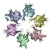
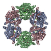
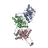
| Title | Hexameric coxsackievirus B3 2C protein in complex with S-fluoxetine | ||||||||||||||||||
 Map data Map data | Hexameric coxsackievirus B3 2C protein in complex with S-fluoxetine | ||||||||||||||||||
 Sample Sample |
| ||||||||||||||||||
| Biological species |  Coxsackievirus B3 (strain Nancy) Coxsackievirus B3 (strain Nancy) | ||||||||||||||||||
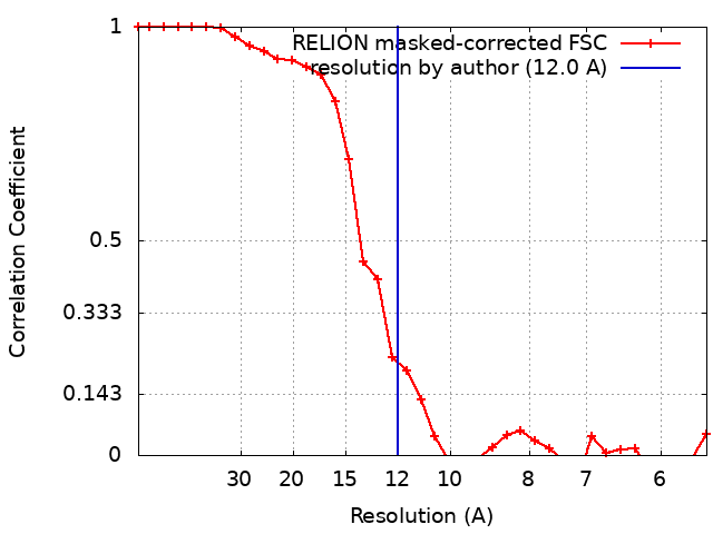
| Method | single particle reconstruction / cryo EM / Resolution: 12.0 Å | ||||||||||||||||||
 Authors Authors | Hurdiss DL / Forster F | ||||||||||||||||||
| Funding support |  Netherlands, 5 items Netherlands, 5 items
| ||||||||||||||||||
 Citation Citation | Journal: Sci Adv / Year: 2022 Title: Fluoxetine targets an allosteric site in the enterovirus 2C AAA+ ATPase and stabilizes a ring-shaped hexameric complex. Authors: Daniel L Hurdiss / Priscila El Kazzi / Lisa Bauer / Nicolas Papageorgiou / François P Ferron / Tim Donselaar / Arno L W van Vliet / Tatiana M Shamorkina / Joost Snijder / Bruno Canard / ...Authors: Daniel L Hurdiss / Priscila El Kazzi / Lisa Bauer / Nicolas Papageorgiou / François P Ferron / Tim Donselaar / Arno L W van Vliet / Tatiana M Shamorkina / Joost Snijder / Bruno Canard / Etienne Decroly / Andrea Brancale / Tzviya Zeev-Ben-Mordehai / Friedrich Förster / Frank J M van Kuppeveld / Bruno Coutard /   Abstract: Enteroviruses are globally prevalent human pathogens responsible for many diseases. The nonstructural protein 2C is a AAA+ helicase and plays a key role in enterovirus replication. Drug repurposing ...Enteroviruses are globally prevalent human pathogens responsible for many diseases. The nonstructural protein 2C is a AAA+ helicase and plays a key role in enterovirus replication. Drug repurposing screens identified 2C-targeting compounds such as fluoxetine and dibucaine, but how they inhibit 2C is unknown. Here, we present a crystal structure of the soluble and monomeric fragment of coxsackievirus B3 2C protein in complex with ()-fluoxetine (SFX), revealing an allosteric binding site. To study the functional consequences of SFX binding, we engineered an adenosine triphosphatase (ATPase)–competent, hexameric 2C protein. Using this system, we show that SFX, dibucaine, HBB [2-(α-hydroxybenzyl)-benzimidazole], and guanidine hydrochloride inhibit 2C ATPase activity. Moreover, cryo–electron microscopy analysis demonstrated that SFX and dibucaine lock 2C in a defined hexameric state, rationalizing their mode of inhibition. Collectively, these results provide important insights into 2C inhibition and a robust engineering strategy for structural, functional, and drug-screening analysis of 2C proteins. | ||||||||||||||||||
| History |
|
- Structure visualization
| Movie |
 Movie viewer Movie viewer |
|---|---|
| Structure viewer | EM map: SurfViewMolmilJmol/JSmol |
| Supplemental images |

- Downloads & links
-EMDB archive
| Map data | emd_12798.map.gz | 203.8 KB | EMDB map data format | |
|---|---|---|---|---|
| Header (meta data) | emd-12798-v30.xmlemd-12798.xml | 18.4 KB 18.4 KB | Display Display | EMDB header |
| FSC (resolution estimation) | emd_12798_fsc.xml | 3 KB | Display | FSC data file |
| Images | 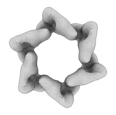 emd_12798.png emd_12798.png | 54.9 KB | ||
| Others | emd_12798_half_map_1.map.gzemd_12798_half_map_2.map.gz | 1.1 MB 1.1 MB | ||
| Archive directory |  http://ftp.pdbj.org/pub/emdb/structures/EMD-12798ftp://ftp.pdbj.org/pub/emdb/structures/EMD-12798 http://ftp.pdbj.org/pub/emdb/structures/EMD-12798ftp://ftp.pdbj.org/pub/emdb/structures/EMD-12798 | HTTPS FTP |
-Related structure data


-Links
| EMDB pages | EMDB (EBI/PDBe) / EMDataResource |
|---|
-Map
| File | Download / File: emd_12798.map.gz / Format: CCP4 / Size: 2 MB / Type: IMAGE STORED AS FLOATING POINT NUMBER (4 BYTES) | ||||||||||||||||||||||||||||||||||||||||||||||||||||||||||||
|---|---|---|---|---|---|---|---|---|---|---|---|---|---|---|---|---|---|---|---|---|---|---|---|---|---|---|---|---|---|---|---|---|---|---|---|---|---|---|---|---|---|---|---|---|---|---|---|---|---|---|---|---|---|---|---|---|---|---|---|---|---|
| Annotation | Hexameric coxsackievirus B3 2C protein in complex with S-fluoxetine | ||||||||||||||||||||||||||||||||||||||||||||||||||||||||||||
| Projections & slices | Image control
Images are generated by Spider. | ||||||||||||||||||||||||||||||||||||||||||||||||||||||||||||
| Voxel size | X=Y=Z: 2.76 Å | ||||||||||||||||||||||||||||||||||||||||||||||||||||||||||||
| Density |
| ||||||||||||||||||||||||||||||||||||||||||||||||||||||||||||
| Symmetry | Space group: 1 | ||||||||||||||||||||||||||||||||||||||||||||||||||||||||||||
| Details | EMDB XML:
CCP4 map header:
| ||||||||||||||||||||||||||||||||||||||||||||||||||||||||||||
 Z (Sec.)
 Y (Row.)
 X (Col.)


-Supplemental data
-Half map: Half map 1
| File | emd_12798_half_map_1.map | ||||||||||||
|---|---|---|---|---|---|---|---|---|---|---|---|---|---|
| Annotation | Half map 1 | ||||||||||||
| Projections & Slices |
| ||||||||||||
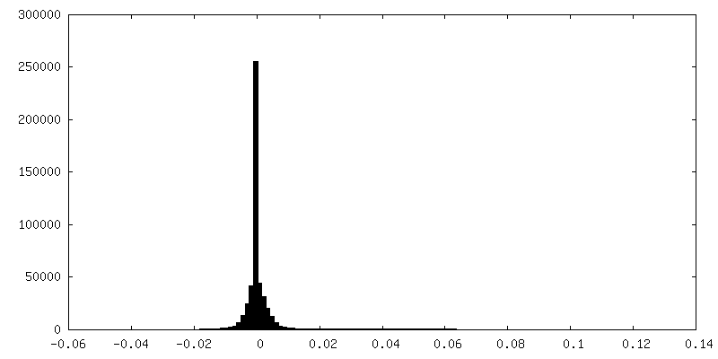
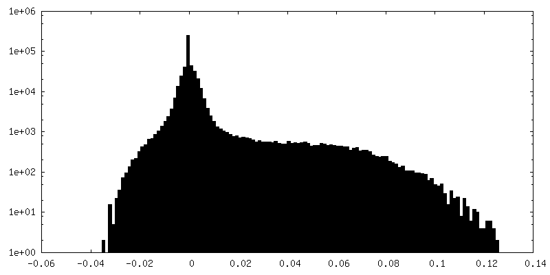
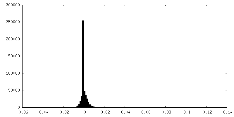
| Density Histograms |


-Half map: Half map 2
| File | emd_12798_half_map_2.map | ||||||||||||
|---|---|---|---|---|---|---|---|---|---|---|---|---|---|
| Annotation | Half map 2 | ||||||||||||
| Projections & Slices |
| ||||||||||||
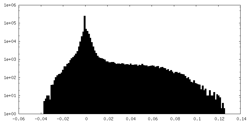
| Density Histograms |


- Sample components
-Entire : Hexameric coxsackievirus B3 2C protein in complex with S-fluoxetine
| Entire | Name: Hexameric coxsackievirus B3 2C protein in complex with S-fluoxetine |
|---|---|
| Components |
|
-Supramolecule #1: Hexameric coxsackievirus B3 2C protein in complex with S-fluoxetine
| Supramolecule | Name: Hexameric coxsackievirus B3 2C protein in complex with S-fluoxetine type: complex / ID: 1 / Parent: 0 / Macromolecule list: all |
|---|---|
| Source (natural) | Organism: Coxsackievirus B3 (strain Nancy) |
| Recombinant expression | Organism:  |
| Molecular weight | Experimental: 170 KDa |
-Macromolecule #1: Coxsackievirus B3 2C protein
| Macromolecule | Name: Coxsackievirus B3 2C protein / type: protein_or_peptide / ID: 1 / Enantiomer: LEVO / EC number: nucleoside-triphosphate phosphatase |
|---|---|
| Source (natural) | Organism: Coxsackievirus B3 (strain Nancy) |
| Recombinant expression | Organism: |
| Sequence | String: GPGGGGSGGG GSGELKAIAQ ELKAIAKELK AIAWELKAIA QGAGGSGSYF QSNASKCRIE PVCLLLHGSP GAGKSVATNL IGRSLAEKLN SSVYSLPPDP DHFDGYKQQA VVIMDDLCQN PDGKDVSLFC QMVSSVDFVP PMAALEEKGI LFTSPFVLAS TNAGSINAPT ...String: GPGGGGSGGG GSGELKAIAQ ELKAIAKELK AIAWELKAIA QGAGGSGSYF QSNASKCRIE PVCLLLHGSP GAGKSVATNL IGRSLAEKLN SSVYSLPPDP DHFDGYKQQA VVIMDDLCQN PDGKDVSLFC QMVSSVDFVP PMAALEEKGI LFTSPFVLAS TNAGSINAPT VSDSRALARR FHFDMNIEVI SMYSQNGKIN MPMSVKTCDD ECCPVNFKKC CPLVCGKAIQ FIDRRTQVRY SLDMLVTEMF REYNHRHSVG TTLEALFQ |
-Experimental details
-Structure determination
| Method | cryo EM |
|---|---|
 Processing Processing | single particle reconstruction |
| Aggregation state | particle |
-Sample preparation
| Concentration | 1 mg/mL | ||||||||||||||||||
|---|---|---|---|---|---|---|---|---|---|---|---|---|---|---|---|---|---|---|---|
| Buffer | pH: 8 Component:
| ||||||||||||||||||
| Grid | Model: Quantifoil R1.2/1.3 / Material: COPPER / Mesh: 200 / Pretreatment - Type: GLOW DISCHARGE / Details: 20 mA using a PELCO easyGLow | ||||||||||||||||||
| Vitrification | Cryogen name: ETHANE / Chamber humidity: 95 % / Chamber temperature: 277 K / Instrument: FEI VITROBOT MARK IV |
- Electron microscopy
| Microscope | TFS KRIOS |
|---|---|
| Details | To account for the preferred orientation exhibited by the sample an alpha tilt of +30 degrees was used for this data collection. |
| Image recording | Film or detector model: GATAN K3 BIOQUANTUM (6k x 4k) / Number grids imaged: 1 / Number real images: 6119 / Average exposure time: 4.0 sec. / Average electron dose: 54.0 e/Å2 |
| Electron beam | Acceleration voltage: 300 kV / Electron source:  FIELD EMISSION GUN FIELD EMISSION GUN |
| Electron optics | Illumination mode: FLOOD BEAM / Imaging mode: BRIGHT FIELD / Cs: 2.7 mm / Nominal defocus max: 4.0 µm / Nominal defocus min: 2.0 µm / Nominal magnification: 64000 |
| Sample stage | Specimen holder model: FEI TITAN KRIOS AUTOGRID HOLDER / Cooling holder cryogen: NITROGEN |
| Experimental equipment |  Model: Titan Krios / Image courtesy: FEI Company |