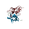
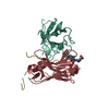
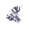
Journal: Sci Adv / Year: 2022 Title: Fluoxetine targets an allosteric site in the enterovirus 2C AAA+ ATPase and stabilizes a ring-shaped hexameric complex. Authors: Daniel L Hurdiss / Priscila El Kazzi / Lisa Bauer / Nicolas Papageorgiou / François P Ferron / Tim Donselaar / Arno L W van Vliet / Tatiana M Shamorkina / Joost Snijder / Bruno Canard / ...Authors: Daniel L Hurdiss / Priscila El Kazzi / Lisa Bauer / Nicolas Papageorgiou / François P Ferron / Tim Donselaar / Arno L W van Vliet / Tatiana M Shamorkina / Joost Snijder / Bruno Canard / Etienne Decroly / Andrea Brancale / Tzviya Zeev-Ben-Mordehai / Friedrich Förster / Frank J M van Kuppeveld / Bruno Coutard / Abstract: Enteroviruses are globally prevalent human pathogens responsible for many diseases. The nonstructural protein 2C is a AAA+ helicase and plays a key role in enterovirus replication. Drug repurposing ...Enteroviruses are globally prevalent human pathogens responsible for many diseases. The nonstructural protein 2C is a AAA+ helicase and plays a key role in enterovirus replication. Drug repurposing screens identified 2C-targeting compounds such as fluoxetine and dibucaine, but how they inhibit 2C is unknown. Here, we present a crystal structure of the soluble and monomeric fragment of coxsackievirus B3 2C protein in complex with ()-fluoxetine (SFX), revealing an allosteric binding site. To study the functional consequences of SFX binding, we engineered an adenosine triphosphatase (ATPase)–competent, hexameric 2C protein. Using this system, we show that SFX, dibucaine, HBB [2-(α-hydroxybenzyl)-benzimidazole], and guanidine hydrochloride inhibit 2C ATPase activity. Moreover, cryo–electron microscopy analysis demonstrated that SFX and dibucaine lock 2C in a defined hexameric state, rationalizing their mode of inhibition. Collectively, these results provide important insights into 2C inhibition and a robust engineering strategy for structural, functional, and drug-screening analysis of 2C proteins.
In the structure databanks used in Yorodumi, some data are registered as the other names, "COVID-19 virus" and "2019-nCoV". Here are the details of the virus and the list of structure data.
Jan 31, 2019. EMDB accession codes are about to change! (news from PDBe EMDB page)
EMDB accession codes are about to change! (news from PDBe EMDB page)
The allocation of 4 digits for EMDB accession codes will soon come to an end. Whilst these codes will remain in use, new EMDB accession codes will include an additional digit and will expand incrementally as the available range of codes is exhausted. The current 4-digit format prefixed with “EMD-” (i.e. EMD-XXXX) will advance to a 5-digit format (i.e. EMD-XXXXX), and so on. It is currently estimated that the 4-digit codes will be depleted around Spring 2019, at which point the 5-digit format will come into force.
The EM Navigator/Yorodumi systems omit the EMD- prefix.
Related info.:Q: What is EMD? / ID/Accession-code notation in Yorodumi/EM Navigator
Yorodumi is a browser for structure data from EMDB, PDB, SASBDB, etc.
This page is also the successor to EM Navigator detail page, and also detail information page/front-end page for Omokage search.
The word "yorodu" (or yorozu) is an old Japanese word meaning "ten thousand". "mi" (miru) is to see.
Related info.:EMDB / PDB / SASBDB / Comparison of 3 databanks / Yorodumi Search / Aug 31, 2016. New EM Navigator & Yorodumi / Yorodumi Papers / Jmol/JSmol / Function and homology information / Changes in new EM Navigator and Yorodumi
 Movie
Movie Controller


 Open data
 Basic information
 Components
 Keywords
 Function and homology information
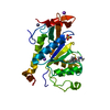
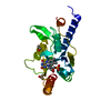
 Coxsackievirus B3
 X-RAY DIFFRACTION /
  Authors
 Citation


 Structure visualization
 Downloads & links
 Other downloads


 PDBj
PDBj

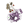
 Assembly

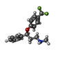
 Mass: 309.326 Da / Num. of mol.: 1 / Source method: obtained synthetically / Formula: C17H18F3NO / Feature type: SUBJECT OF INVESTIGATION
 Mass: 65.409 Da / Num. of mol.: 1 / Source method: obtained synthetically / Formula: Zn
 Mass: 35.453 Da / Num. of mol.: 1 / Source method: obtained synthetically / Formula: Cl
 Mass: 18.015 Da / Num. of mol.: 211 / Source method: isolated from a natural source / Formula: H2O
 Sample preparation
 Processing