 Movie
Movie Controller

[English] 日本語
 Yorodumi
- PDB-1nlf: Crystal Structure of DNA Helicase RepA in complex with sulfate at... -
+ Open data

- Basic information
| Entry | Database: PDB / ID: 1nlf | ||||||
|---|---|---|---|---|---|---|---|
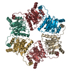
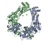
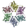
| Title | Crystal Structure of DNA Helicase RepA in complex with sulfate at 1.95 A resolution | ||||||
 Components Components | Regulatory protein repA | ||||||
 Keywords Keywords | REPLICATION / replicative DNA helicase structural changes | ||||||
| Function / homology |  Function and homology information Function and homology information | ||||||
| Biological species |  | ||||||
| Method |  X-RAY DIFFRACTION / SYNCHROTRON / MOLECULAR REPLACEMENT / Resolution: 1.95 Å X-RAY DIFFRACTION / SYNCHROTRON / MOLECULAR REPLACEMENT / Resolution: 1.95 Å | ||||||
 Authors Authors | Xu, H. / Strater, N. / Schroeder, W. / Bottcher, C. / Ludwig, K. / Saenger, W. | ||||||
 Citation Citation | Journal: Acta Crystallogr.,Sect.D / Year: 2003 Title: Structure of DNA helicase RepA in complex with sulfate at 1.95 A resolution implicates structural changes to an "open" form. Authors: Xu, H. / Strater, N. / Schroder, W. / Bottcher, C. / Ludwig, K. / Saenger, W. | ||||||
| History |
|
- Structure visualization
| Structure viewer | Molecule: MolmilJmol/JSmol |
|---|
- Downloads & links
-Download
| PDBx/mmCIF format | 1nlf.cif.gz | 170.2 KB | Display | PDBx/mmCIF format |
|---|---|---|---|---|
| PDB format | pdb1nlf.ent.gz | 134.4 KB | Display | PDB format |
| PDBx/mmJSON format | 1nlf.json.gz | Tree view | PDBx/mmJSON format | |
| Others |  Other downloads Other downloads |
-Validation report
| Arichive directory | https://data.pdbj.org/pub/pdb/validation_reports/nl/1nlfftp://data.pdbj.org/pub/pdb/validation_reports/nl/1nlf | HTTPS FTP |
|---|
-Related structure data
| Similar structure data |
|---|


-Links
 PDBj
PDBj- Assembly
| Deposited unit | 
| ||||||||
|---|---|---|---|---|---|---|---|---|---|
| 1 | 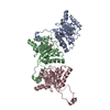
| ||||||||
| Unit cell |
| ||||||||
| Details | The second part of the biological assembly is generated by the crystallographic 2-fold rotation axes parallel to b and stacked face-to-face in a and c directions. |
-Components
| #1: Protein | Mass: 29945.189 Da / Num. of mol.: 3 Source method: isolated from a genetically manipulated source Source: (gene. exp.) #2: Chemical |   Mass: 96.063 Da / Num. of mol.: 3 / Source method: obtained synthetically / Formula: SO4 Mass: 96.063 Da / Num. of mol.: 3 / Source method: obtained synthetically / Formula: SO4#3: Water | ChemComp-HOH / |  Mass: 18.015 Da / Num. of mol.: 756 / Source method: isolated from a natural source / Formula: H2O Mass: 18.015 Da / Num. of mol.: 756 / Source method: isolated from a natural source / Formula: H2O |
|---|
-Experimental details
-Experiment
| Experiment | Method: X-RAY DIFFRACTION / Number of used crystals: 1 |
|---|
- Sample preparation
| Crystal | Density Matthews: 2.56 Å3/Da / Density % sol: 51.58 % | ||||||||||||||||||||||||||||||||||||||||||||||||||||||||||||||||||||||
|---|---|---|---|---|---|---|---|---|---|---|---|---|---|---|---|---|---|---|---|---|---|---|---|---|---|---|---|---|---|---|---|---|---|---|---|---|---|---|---|---|---|---|---|---|---|---|---|---|---|---|---|---|---|---|---|---|---|---|---|---|---|---|---|---|---|---|---|---|---|---|---|
| Crystal grow | Temperature: 291 K / Method: vapor diffusion, hanging drop / pH: 8 Details: PEG 4000, TRIS buffer, magnesium sulfate , pH 8.0, VAPOR DIFFUSION, HANGING DROP, temperature 291K | ||||||||||||||||||||||||||||||||||||||||||||||||||||||||||||||||||||||
| Crystal grow | *PLUS Temperature: 277 K / pH: 5.8 | ||||||||||||||||||||||||||||||||||||||||||||||||||||||||||||||||||||||
| Components of the solutions | *PLUS
|
-Data collection
| Diffraction | Mean temperature: 100 K |
|---|---|
| Diffraction source | Source: SYNCHROTRON / Site: BESSY  / Beamline: 14.2 / Wavelength: 0.83432 Å / Beamline: 14.2 / Wavelength: 0.83432 Å |
| Detector | Type: MARRESEARCH / Detector: IMAGE PLATE / Date: May 18, 2001 |
| Radiation | Protocol: SINGLE WAVELENGTH / Monochromatic (M) / Laue (L): M / Scattering type: x-ray |
| Radiation wavelength | Wavelength: 0.83432 Å / Relative weight: 1 |
| Reflection | Resolution: 1.95→20 Å / Num. all: 87437 / Num. obs: 72794 / % possible obs: 98.4 % / Observed criterion σ(F): 0 / Observed criterion σ(I): 0 / Redundancy: 3.3 % / Biso Wilson estimate: 21.4 Å2 / Rmerge(I) obs: 0.078 / Net I/σ(I): 9.1 |
| Reflection | *PLUS Lowest resolution: 20 Å / Num. measured all: 243785 |
| Reflection shell | *PLUS % possible obs: 99.6 % / Rmerge(I) obs: 0.097 / Mean I/σ(I) obs: 6.8 |
- Processing
| Software |
| |||||||||||||||||||||||||
|---|---|---|---|---|---|---|---|---|---|---|---|---|---|---|---|---|---|---|---|---|---|---|---|---|---|---|
| Refinement | Method to determine structure: MOLECULAR REPLACEMENT / Resolution: 1.95→20 Å / Isotropic thermal model: Isotropic / σ(F): 2 / Stereochemistry target values: Engh & Huber
| |||||||||||||||||||||||||
| Displacement parameters | Biso mean: 21.4 Å2 | |||||||||||||||||||||||||
| Refinement step | Cycle: LAST / Resolution: 1.95→20 Å
| |||||||||||||||||||||||||
| Refine LS restraints |
| |||||||||||||||||||||||||
| Refinement | *PLUS Lowest resolution: 20 Å / % reflection Rfree: 5 % | |||||||||||||||||||||||||
| Solvent computation | *PLUS | |||||||||||||||||||||||||
| Displacement parameters | *PLUS |
