 ムービー
ムービー コントローラー
 構造ビューア
 EMN検索について


-検索条件
-検索結果
検索 (著者・登録者: altman & r)の結果64件中、1から50件目までを表示しています
EMDB-29757: 
mRNA decoding in human is kinetically and structurally distinct from bacteria (IC state)

EMDB-29758:
mRNA decoding in human is kinetically and structurally distinct from bacteria (GA state)

EMDB-29759:
mRNA decoding in human is kinetically and structurally distinct from bacteria (CR state)
EMDB-29760:
mRNA decoding in human is kinetically and structurally distinct from bacteria (AC state)

EMDB-29766:
mRNA decoding in human is kinetically and structurally distinct from bacteria (60S Focus refined map)

EMDB-29768:
mRNA decoding in human is kinetically and structurally distinct from bacteria (40S Focus refined map)

EMDB-29771:
mRNA decoding in human is kinetically and structurally distinct from bacteria (GA state 2)

EMDB-29782:
mRNA decoding in human is kinetically and structurally distinct from bacteria (80S consensus refined structure)
EMDB-40205:
mRNA decoding in human is kinetically and structurally distinct from bacteria (Consensus LSU focused refined structure)
PDB-8g5y:
mRNA decoding in human is kinetically and structurally distinct from bacteria (IC state)

PDB-8g5z:
mRNA decoding in human is kinetically and structurally distinct from bacteria (GA state)

PDB-8g60:
mRNA decoding in human is kinetically and structurally distinct from bacteria (CR state)
PDB-8g61:
mRNA decoding in human is kinetically and structurally distinct from bacteria (AC state)

PDB-8g6j:
mRNA decoding in human is kinetically and structurally distinct from bacteria (GA state 2)
PDB-8glp:
mRNA decoding in human is kinetically and structurally distinct from bacteria (Consensus LSU focused refined structure)
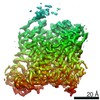
EMDB-24120:
Elongating 70S ribosome complex in a classical pre-translocation (PRE-C) conformation
EMDB-24132:
Elongating 70S ribosome complex in a fusidic acid-stalled intermediate state of translocation bound to EF-G(GDP) (INT2)
EMDB-24133:
Elongating 70S ribosome complex in a hybrid-H1 pre-translocation (PRE-H1) conformation
EMDB-24134:
Elongating 70S ribosome complex in a spectinomycin-stalled intermediate state of translocation bound to EF-G in an active, GTP conformation (INT1)
EMDB-24135:
Elongating 70S ribosome complex in a hybrid-H2* pre-translocation (PRE-H2*) conformation
EMDB-24136:
Elongating 70S ribosome complex in a post-translocation (POST) conformation
PDB-7n1p:
Elongating 70S ribosome complex in a classical pre-translocation (PRE-C) conformation
PDB-7n2c:
Elongating 70S ribosome complex in a fusidic acid-stalled intermediate state of translocation bound to EF-G(GDP) (INT2)
PDB-7n2u:
Elongating 70S ribosome complex in a hybrid-H1 pre-translocation (PRE-H1) conformation
PDB-7n2v:
Elongating 70S ribosome complex in a spectinomycin-stalled intermediate state of translocation bound to EF-G in an active, GTP conformation (INT1)
PDB-7n30:
Elongating 70S ribosome complex in a hybrid-H2* pre-translocation (PRE-H2*) conformation
PDB-7n31:
Elongating 70S ribosome complex in a post-translocation (POST) conformation

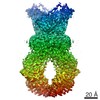
EMDB-30835:
cryo EM map of the LAT1-4F2hc bound with JX-075

EMDB-30836:
cryo EM map of the LAT1-4F2hc bound with JX-075, focused refined on transmembrane region

EMDB-30837:
cryo EM map of the LAT1-4F2hc bound with JX-078
EMDB-30838:
cryo EM map of the LAT1-4F2hc bound with JX-078, focused refined on transmembrane region

EMDB-30839:
cryo EM map of the LAT1-4F2hc bound with JX-119

EMDB-30840:
cryo EM map of the LAT1-4F2hc bound with JX-119, focused refined on transmembrane region

EMDB-30841:
cryo EM map of the LAT1-4F2hc bound with 3,5-diiodo-L-tyrosine

EMDB-30842:
cryo EM map of the LAT1-4F2hc bound with 3,5-diiodo-L-tyrosine, focused refined on transmembrane region

PDB-7dsk:
Overall structure of the LAT1-4F2hc bound with JX-075

PDB-7dsl:
Overall structure of the LAT1-4F2hc bound with JX-078

PDB-7dsn:
Overall structure of the LAT1-4F2hc bound with JX-119

PDB-7dsq:
Overall structure of the LAT1-4F2hc bound with 3,5-diiodo-L-tyrosine

EMDB-20293:
EBOV GPdTM (Mayinga) in complex with rEBOV-548 Fab

EMDB-20301:
EBOV GPdMuc (Makona) in complex with rEBOV-520 and rEBOV-548 Fabs

EMDB-20947:
EBOV GPdMuc Makona bound to rEBOV-548 Fab

PDB-6pci:
EBOV GPdMuc (Makona) in complex with rEBOV-520 and rEBOV-548 Fabs

PDB-6uye:
EBOV GPdMuc Makona bound to rEBOV-548 Fab

PDB-6hij:
Cryo-EM structure of the human ABCG2-MZ29-Fab complex with cholesterol and PE lipids docked
EMDB-3953:
Structure of inhibitor-bound ABCG2
EMDB-4246:
Structure of inhibitor-bound ABCG2
EMDB-4256:
Structure of an inhibitor-bound ABC transporter

PDB-6eti:
Structure of inhibitor-bound ABCG2

PDB-6feq:
Structure of inhibitor-bound ABCG2
ページ:
 wwPDBはEMDBデータモデルのバージョン3へ移行します
