Movie
Movie Controller

[English] 日本語
 Yorodumi
- EMDB-20301: EBOV GPdMuc (Makona) in complex with rEBOV-520 and rEBOV-548 Fabs -
+ Open data

- Basic information
| Entry | Database: EMDB / ID: EMD-20301 | |||||||||
|---|---|---|---|---|---|---|---|---|---|---|
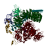
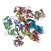
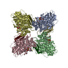
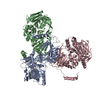
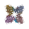
| Title | EBOV GPdMuc (Makona) in complex with rEBOV-520 and rEBOV-548 Fabs | |||||||||
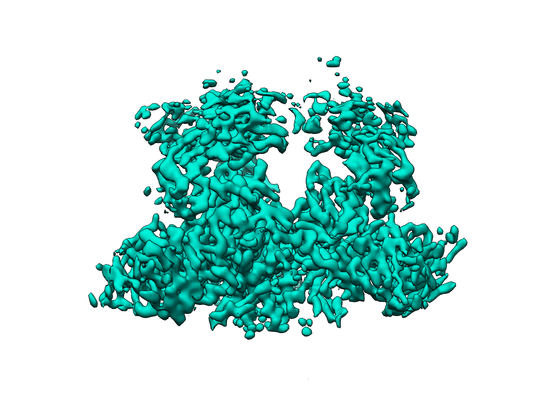
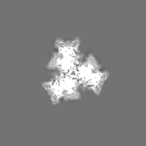
 Map data Map data | EBOVGPdMuc (Makona) in complex with rEBOV-520 and rEBOV-548 Fabs | |||||||||
 Sample Sample |
| |||||||||
 Keywords Keywords | Ebola / filovirus / antibody / synergy / IMMUNE SYSTEM / viral protein | |||||||||
| Function / homology |  Function and homology information Function and homology informationsymbiont-mediated-mediated suppression of host tetherin activity / clathrin-dependent endocytosis of virus by host cell / entry receptor-mediated virion attachment to host cell / symbiont-mediated suppression of host innate immune response / fusion of virus membrane with host endosome membrane / viral envelope / lipid binding / host cell plasma membrane / virion membrane / extracellular region Similarity search - Function | |||||||||
| Biological species |  Homo sapiens (human) / Homo sapiens (human) /   Ebola virus Ebola virus | |||||||||
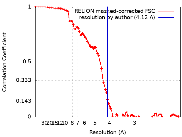
| Method | single particle reconstruction / cryo EM / Resolution: 4.12 Å | |||||||||
 Authors Authors | Ward AB / Murin CD | |||||||||
| Funding support |  United States, 1 items United States, 1 items
| |||||||||
 Citation Citation | Journal: Immunity / Year: 2020 Title: Analysis of a Therapeutic Antibody Cocktail Reveals Determinants for Cooperative and Broad Ebolavirus Neutralization. Authors: Pavlo Gilchuk / Charles D Murin / Jacob C Milligan / Robert W Cross / Chad E Mire / Philipp A Ilinykh / Kai Huang / Natalia Kuzmina / Pilar X Altman / Sean Hui / Bronwyn M Gunn / Aubrey L ...Authors: Pavlo Gilchuk / Charles D Murin / Jacob C Milligan / Robert W Cross / Chad E Mire / Philipp A Ilinykh / Kai Huang / Natalia Kuzmina / Pilar X Altman / Sean Hui / Bronwyn M Gunn / Aubrey L Bryan / Edgar Davidson / Benjamin J Doranz / Hannah L Turner / Tanwee Alkutkar / Robin Flinko / Chiara Orlandi / Robert Carnahan / Rachel Nargi / Robin G Bombardi / Megan E Vodzak / Sheng Li / Adaora Okoli / Morris Ibeawuchi / Benjamin Ohiaeri / George K Lewis / Galit Alter / Alexander Bukreyev / Erica Ollmann Saphire / Thomas W Geisbert / Andrew B Ward / James E Crowe / Abstract: Structural principles underlying the composition of protective antiviral monoclonal antibody (mAb) cocktails are poorly defined. Here, we exploited antibody cooperativity to develop a therapeutic mAb ...Structural principles underlying the composition of protective antiviral monoclonal antibody (mAb) cocktails are poorly defined. Here, we exploited antibody cooperativity to develop a therapeutic mAb cocktail against Ebola virus. We systematically analyzed the antibody repertoire in human survivors and identified a pair of potently neutralizing mAbs that cooperatively bound to the ebolavirus glycoprotein (GP). High-resolution structures revealed that in a two-antibody cocktail, molecular mimicry was a major feature of mAb-GP interactions. Broadly neutralizing mAb rEBOV-520 targeted a conserved epitope on the GP base region. mAb rEBOV-548 bound to a glycan cap epitope, possessed neutralizing and Fc-mediated effector function activities, and potentiated neutralization by rEBOV-520. Remodeling of the glycan cap structures by the cocktail enabled enhanced GP binding and virus neutralization. The cocktail demonstrated resistance to virus escape and protected non-human primates (NHPs) against Ebola virus disease. These data illuminate structural principles of antibody cooperativity with implications for development of antiviral immunotherapeutics. | |||||||||
| History |
|
- Structure visualization
| Movie |
Movie viewer |
|---|---|
| Structure viewer | EM map: SurfViewMolmilJmol/JSmol |
| Supplemental images |
- Downloads & links
-EMDB archive
| Map data | emd_20301.map.gz | 7.5 MB | EMDB map data format | |
|---|---|---|---|---|
| Header (meta data) | emd-20301-v30.xmlemd-20301.xml | 22.8 KB 22.8 KB | Display Display | EMDB header |
| FSC (resolution estimation) | emd_20301_fsc.xml | 10.3 KB | Display | FSC data file |
| Images |  emd_20301.png emd_20301.png | 143.4 KB | ||
| Filedesc metadata | emd-20301.cif.gz | 7.4 KB | ||
| Archive directory |  http://ftp.pdbj.org/pub/emdb/structures/EMD-20301ftp://ftp.pdbj.org/pub/emdb/structures/EMD-20301 http://ftp.pdbj.org/pub/emdb/structures/EMD-20301ftp://ftp.pdbj.org/pub/emdb/structures/EMD-20301 | HTTPS FTP |
-Related structure data
| Related structure data |  6pciMC 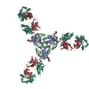 6oz9C  6uyeC M: atomic model generated by this map C: citing same article ( |
|---|---|
| Similar structure data |


-Links
| EMDB pages | EMDB (EBI/PDBe) / EMDataResource |
|---|---|
| Related items in Molecule of the Month |


-Map
| File | Download / File: emd_20301.map.gz / Format: CCP4 / Size: 91.1 MB / Type: IMAGE STORED AS FLOATING POINT NUMBER (4 BYTES) | ||||||||||||||||||||||||||||||||||||||||||||||||||||||||||||||||||||
|---|---|---|---|---|---|---|---|---|---|---|---|---|---|---|---|---|---|---|---|---|---|---|---|---|---|---|---|---|---|---|---|---|---|---|---|---|---|---|---|---|---|---|---|---|---|---|---|---|---|---|---|---|---|---|---|---|---|---|---|---|---|---|---|---|---|---|---|---|---|
| Annotation | EBOVGPdMuc (Makona) in complex with rEBOV-520 and rEBOV-548 Fabs | ||||||||||||||||||||||||||||||||||||||||||||||||||||||||||||||||||||
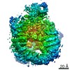
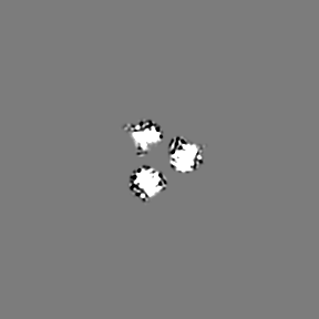
| Projections & slices | Image control
Images are generated by Spider. | ||||||||||||||||||||||||||||||||||||||||||||||||||||||||||||||||||||
| Voxel size | X=Y=Z: 1.03 Å | ||||||||||||||||||||||||||||||||||||||||||||||||||||||||||||||||||||
| Density |
| ||||||||||||||||||||||||||||||||||||||||||||||||||||||||||||||||||||
| Symmetry | Space group: 1 | ||||||||||||||||||||||||||||||||||||||||||||||||||||||||||||||||||||
| Details | EMDB XML:
CCP4 map header:
| ||||||||||||||||||||||||||||||||||||||||||||||||||||||||||||||||||||
 Z (Sec.)
 Y (Row.)
 X (Col.)


-Supplemental data
- Sample components
+Entire : EBOV GPdMuc (Makona) in complex with rEBOV-520 and rEBOV-548 Fabs
+Supramolecule #1: EBOV GPdMuc (Makona) in complex with rEBOV-520 and rEBOV-548 Fabs
+Supramolecule #2: Ebola Virus (Makona) GP
+Supramolecule #3: rEBOV-548 Fab
+Supramolecule #4: rEBOV-520 Fab
+Macromolecule #1: Virion spike glycoprotein
+Macromolecule #2: rEBOV-548 Fab heavy chain
+Macromolecule #3: rEBOV-548 Fab light chain
+Macromolecule #4: Virion spike glycoprotein,Virion spike glycoprotein,Ebola Virus (...
+Macromolecule #5: rEBOV-520 Fab light chain
+Macromolecule #6: rEBOV-520 Fab heavy chain
+Macromolecule #8: 2-acetamido-2-deoxy-beta-D-glucopyranose

-Experimental details
-Structure determination
| Method | cryo EM |
|---|---|
 Processing Processing | single particle reconstruction |
| Aggregation state | particle |
-Sample preparation
| Concentration | 4 mg/mL | ||||||||||||
|---|---|---|---|---|---|---|---|---|---|---|---|---|---|
| Buffer | pH: 7.2 Component:
Details: Buffers were made fresh from 10X stocks. Detergent was made fresh in TBS as a 6X stock and added immediately prior to vitificaiton. | ||||||||||||
| Grid | Model: Quantifoil R1.2/1.3 / Material: COPPER / Mesh: 400 / Support film - Material: CARBON / Support film - topology: HOLEY | ||||||||||||
| Vitrification | Cryogen name: ETHANE / Chamber humidity: 100 % / Chamber temperature: 277.15 K / Instrument: FEI VITROBOT MARK IV Details: Sample was blotted on both sides of the grid. Sample equilibrated in the chamber for 10s before blotting for 5s.. |
- Electron microscopy
| Microscope | FEI TITAN KRIOS |
|---|---|
| Image recording | Film or detector model: GATAN K2 SUMMIT (4k x 4k) / Detector mode: COUNTING / Number grids imaged: 1 / Number real images: 2832 / Average exposure time: 9.5 sec. / Average electron dose: 51.85 e/Å2 |
| Electron beam | Acceleration voltage: 300 kV / Electron source:  FIELD EMISSION GUN FIELD EMISSION GUN |
| Electron optics | C2 aperture diameter: 70.0 µm / Illumination mode: FLOOD BEAM / Imaging mode: BRIGHT FIELD / Cs: 2.7 mm / Nominal defocus max: 3.0 µm / Nominal defocus min: 0.5 µm / Nominal magnification: 29000 |
| Sample stage | Specimen holder model: FEI TITAN KRIOS AUTOGRID HOLDER / Cooling holder cryogen: NITROGEN |
| Experimental equipment |  Model: Titan Krios / Image courtesy: FEI Company |