 Movie
Movie Controller


+ Open data

- Basic information
| Entry | Database: PDB / ID: 5jq3 | |||||||||
|---|---|---|---|---|---|---|---|---|---|---|
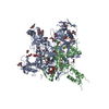
| Title | Crystal structure of Ebola glycoprotein | |||||||||
 Components Components | (Envelope glycoprotein ...) x 2 | |||||||||
 Keywords Keywords | VIRAL PROTEIN / Ebola virus / Filoviridae / envelope glycoprotein / protein inhibitor complex / ibuprofen / toremifene | |||||||||
| Function / homology |  Function and homology information Function and homology informationsymbiont-mediated killing of host cell / host cell endoplasmic reticulum / viral budding from plasma membrane / symbiont-mediated-mediated suppression of host tetherin activity / clathrin-dependent endocytosis of virus by host cell / host cell cytoplasm / entry receptor-mediated virion attachment to host cell / symbiont-mediated suppression of host innate immune response / membrane raft / fusion of virus membrane with host endosome membrane ...symbiont-mediated killing of host cell / host cell endoplasmic reticulum / viral budding from plasma membrane / symbiont-mediated-mediated suppression of host tetherin activity / clathrin-dependent endocytosis of virus by host cell / host cell cytoplasm / entry receptor-mediated virion attachment to host cell / symbiont-mediated suppression of host innate immune response / membrane raft / fusion of virus membrane with host endosome membrane / viral envelope / symbiont entry into host cell / lipid binding / host cell plasma membrane / virion membrane / extracellular region / identical protein binding Similarity search - Function | |||||||||
| Biological species |   Ebola virus - MayingaZaire (virus) Ebola virus - MayingaZaire (virus)1976 | |||||||||
| Method |  X-RAY DIFFRACTION / SYNCHROTRON / MOLECULAR REPLACEMENT / Resolution: 2.23 Å X-RAY DIFFRACTION / SYNCHROTRON / MOLECULAR REPLACEMENT / Resolution: 2.23 Å | |||||||||
 Authors Authors | Zhao, Y. / Ren, J. / Stuart, D.I. | |||||||||
 Citation Citation | Journal: Nature / Year: 2016 Title: Toremifene interacts with and destabilizes the Ebola virus glycoprotein. Authors: Zhao, Y. / Ren, J. / Harlos, K. / Jones, D.M. / Zeltina, A. / Bowden, T.A. / Padilla-Parra, S. / Fry, E.E. / Stuart, D.I. | |||||||||
| History |
|
- Structure visualization
| Structure viewer | Molecule: MolmilJmol/JSmol |
|---|
- Downloads & links
-Download
| PDBx/mmCIF format | 5jq3.cif.gz | 179.5 KB | Display | PDBx/mmCIF format |
|---|---|---|---|---|
| PDB format | pdb5jq3.ent.gz | 140.8 KB | Display | PDB format |
| PDBx/mmJSON format | 5jq3.json.gz | Tree view | PDBx/mmJSON format | |
| Others |  Other downloads Other downloads |
-Validation report
| Arichive directory | https://data.pdbj.org/pub/pdb/validation_reports/jq/5jq3ftp://data.pdbj.org/pub/pdb/validation_reports/jq/5jq3 | HTTPS FTP |
|---|
-Related structure data
| Related structure data |  5jq7C 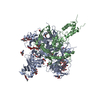 5jqbC  3csyS S: Starting model for refinement C: citing same article ( |
|---|---|
| Similar structure data |


-Links
 PDBj
PDBj

- Assembly
| Deposited unit | 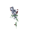
| ||||||||
|---|---|---|---|---|---|---|---|---|---|
| 1 | 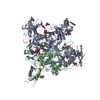
| ||||||||
| Unit cell |
|
-Components
-Envelope glycoprotein ... , 2 types, 2 molecules AB
| #1: Protein | Mass: 36472.930 Da / Num. of mol.: 1 / Mutation: T42A,T42A,T42A Source method: isolated from a genetically manipulated source Source: (gene. exp.) Ebola virus - Mayinga, Zaire, 1976 / Gene: GP / Cell line (production host): HEK293T / Production host:  Homo sapiens (human) / References: UniProt: Q05320 Homo sapiens (human) / References: UniProt: Q05320 |
|---|---|
| #2: Protein | Mass: 18989.391 Da / Num. of mol.: 1 Source method: isolated from a genetically manipulated source Source: (gene. exp.) Ebola virus - Mayinga, Zaire, 1976 / Gene: GP / Cell line (production host): HEK293T / Production host: Homo sapiens (human) / References: UniProt: Q05320 |
-Sugars , 2 types, 6 molecules 
| #3: Polysaccharide | alpha-D-mannopyranose-(1-3)-[alpha-D-mannopyranose-(1-6)]beta-D-mannopyranose-(1-4)-2-acetamido-2- ...alpha-D-mannopyranose-(1-3)-[alpha-D-mannopyranose-(1-6)]beta-D-mannopyranose-(1-4)-2-acetamido-2-deoxy-beta-D-glucopyranose-(1-4)-2-acetamido-2-deoxy-beta-D-glucopyranose Source method: isolated from a genetically manipulated source |
|---|---|
| #4: Sugar | ChemComp-NAG /  Type: D-saccharide, beta linking / Mass: 221.208 Da / Num. of mol.: 5 Type: D-saccharide, beta linking / Mass: 221.208 Da / Num. of mol.: 5Source method: isolated from a genetically manipulated source Formula: C8H15NO6 |
-Non-polymers , 2 types, 121 molecules 

| #5: Chemical |  Mass: 92.094 Da / Num. of mol.: 2 / Source method: obtained synthetically / Formula: C3H8O3 Mass: 92.094 Da / Num. of mol.: 2 / Source method: obtained synthetically / Formula: C3H8O3#6: Water | ChemComp-HOH / | Mass: 18.015 Da / Num. of mol.: 119 / Source method: isolated from a natural source / Formula: H2O |
|---|
-Details
| Has protein modification | Y |
|---|
-Experimental details
-Experiment
| Experiment | Method: X-RAY DIFFRACTION / Number of used crystals: 1 |
|---|
- Sample preparation
| Crystal | Density Matthews: 4.43 Å3/Da / Density % sol: 72.21 % |
|---|---|
| Crystal grow | Temperature: 293 K / Method: vapor diffusion, sitting drop / pH: 5.2 Details: 9% (w/v) PEG 6000 and 0.1 M Sodium citrate tribasic dihydrate |
-Data collection
| Diffraction | Mean temperature: 100 K |
|---|---|
| Diffraction source | Source: SYNCHROTRON / Site: Diamond  / Beamline: I03 / Wavelength: 1.07193 Å / Beamline: I03 / Wavelength: 1.07193 Å |
| Detector | Type: DECTRIS PILATUS3 6M / Detector: PIXEL / Date: Sep 12, 2015 |
| Radiation | Protocol: SINGLE WAVELENGTH / Monochromatic (M) / Laue (L): M / Scattering type: x-ray |
| Radiation wavelength | Wavelength: 1.07193 Å / Relative weight: 1 |
| Reflection | Resolution: 2.23→94.19 Å / Num. obs: 38090 / % possible obs: 100 % / Redundancy: 57.8 % / Rmerge(I) obs: 0.204 / Net I/σ(I): 17.4 |
| Reflection shell | Resolution: 2.23→2.29 Å / Redundancy: 15.4 % / Mean I/σ(I) obs: 1.3 / % possible all: 100 |
- Processing
| Software |
| ||||||||||||||||||||||||||||||||||||||||||||||||||||||||||||||||||||||||||||||||||||||||||||||||||||||||||||||||||||||||||||||||||||||||||||||||||||||||||||||||||||||||||||||||||||||
|---|---|---|---|---|---|---|---|---|---|---|---|---|---|---|---|---|---|---|---|---|---|---|---|---|---|---|---|---|---|---|---|---|---|---|---|---|---|---|---|---|---|---|---|---|---|---|---|---|---|---|---|---|---|---|---|---|---|---|---|---|---|---|---|---|---|---|---|---|---|---|---|---|---|---|---|---|---|---|---|---|---|---|---|---|---|---|---|---|---|---|---|---|---|---|---|---|---|---|---|---|---|---|---|---|---|---|---|---|---|---|---|---|---|---|---|---|---|---|---|---|---|---|---|---|---|---|---|---|---|---|---|---|---|---|---|---|---|---|---|---|---|---|---|---|---|---|---|---|---|---|---|---|---|---|---|---|---|---|---|---|---|---|---|---|---|---|---|---|---|---|---|---|---|---|---|---|---|---|---|---|---|---|---|
| Refinement | Method to determine structure: MOLECULAR REPLACEMENT Starting model: 3CSY Resolution: 2.23→94.19 Å / Cor.coef. Fo:Fc: 0.942 / Cor.coef. Fo:Fc free: 0.934 / SU B: 12.888 / SU ML: 0.156 / Cross valid method: THROUGHOUT / ESU R: 0.193 / ESU R Free: 0.17 / Details: HYDROGENS HAVE BEEN ADDED IN THE RIDING POSITIONS
| ||||||||||||||||||||||||||||||||||||||||||||||||||||||||||||||||||||||||||||||||||||||||||||||||||||||||||||||||||||||||||||||||||||||||||||||||||||||||||||||||||||||||||||||||||||||
| Solvent computation | Ion probe radii: 0.8 Å / Shrinkage radii: 0.8 Å / VDW probe radii: 1.2 Å | ||||||||||||||||||||||||||||||||||||||||||||||||||||||||||||||||||||||||||||||||||||||||||||||||||||||||||||||||||||||||||||||||||||||||||||||||||||||||||||||||||||||||||||||||||||||
| Displacement parameters | Biso mean: 72.48 Å2
| ||||||||||||||||||||||||||||||||||||||||||||||||||||||||||||||||||||||||||||||||||||||||||||||||||||||||||||||||||||||||||||||||||||||||||||||||||||||||||||||||||||||||||||||||||||||
| Refinement step | Cycle: LAST / Resolution: 2.23→94.19 Å
| ||||||||||||||||||||||||||||||||||||||||||||||||||||||||||||||||||||||||||||||||||||||||||||||||||||||||||||||||||||||||||||||||||||||||||||||||||||||||||||||||||||||||||||||||||||||
| Refine LS restraints |
|