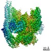
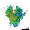
 Movie
Movie Controller


+ Open data

- Basic information
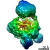
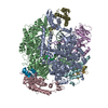
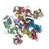
| Entry | Database: PDB / ID: 6y50 | ||||||
|---|---|---|---|---|---|---|---|
| Title | 5'domain of human 17S U2 snRNP | ||||||
 Components Components |
| ||||||
 Keywords Keywords | SPLICING / 17S U2 snRNP | ||||||
| Function / homology |  Function and homology information Function and homology informationU11/U12 snRNP / B-WICH complex / poly-ADP-D-ribose modification-dependent protein binding / U12-type spliceosomal complex / RNA splicing, via transesterification reactions / protein localization to site of double-strand break / splicing factor binding / U2-type precatalytic spliceosome / U2-type prespliceosome assembly / U2-type spliceosomal complex ...U11/U12 snRNP / B-WICH complex / poly-ADP-D-ribose modification-dependent protein binding / U12-type spliceosomal complex / RNA splicing, via transesterification reactions / protein localization to site of double-strand break / splicing factor binding / U2-type precatalytic spliceosome / U2-type prespliceosome assembly / U2-type spliceosomal complex / chromatin-protein adaptor activity / SAGA complex / U2 snRNP / U2-type prespliceosome / positive regulation of transcription by RNA polymerase III / precatalytic spliceosome / mRNA 3'-splice site recognition / regulation of RNA splicing / mRNA Splicing - Minor Pathway / positive regulation of transcription by RNA polymerase I / spliceosomal complex assembly / U2 snRNA binding / Cajal body / regulation of DNA repair / catalytic step 2 spliceosome / mRNA Splicing - Major Pathway / RNA splicing / stem cell differentiation / spliceosomal complex / mRNA splicing, via spliceosome / negative regulation of protein catabolic process / B-WICH complex positively regulates rRNA expression / double-strand break repair via homologous recombination / fibrillar center / nuclear matrix / mRNA processing / site of double-strand break / RNA helicase activity / nuclear speck / RNA helicase / chromatin remodeling / mRNA binding / positive regulation of DNA-templated transcription / protein-containing complex binding / nucleolus / ATP hydrolysis activity / positive regulation of transcription by RNA polymerase II / DNA binding / RNA binding / zinc ion binding / nucleoplasm / ATP binding / nucleus Similarity search - Function | ||||||
| Biological species |  Homo sapiens (human) Homo sapiens (human) | ||||||
| Method | ELECTRON MICROSCOPY / single particle reconstruction / cryo EM / Resolution: 4.1 Å | ||||||
 Authors Authors | Zhang, Z. / Will, C.L. / Bertram, K. / Luehrmann, R. / Stark, H. | ||||||
| Funding support |  Germany, 1items Germany, 1items
| ||||||
 Citation Citation | Journal: Nature / Year: 2020 Title: Molecular architecture of the human 17S U2 snRNP. Authors: Zhenwei Zhang / Cindy L Will / Karl Bertram / Olexandr Dybkov / Klaus Hartmuth / Dmitry E Agafonov / Romina Hofele / Henning Urlaub / Berthold Kastner / Reinhard Lührmann / Holger Stark /  Abstract: The U2 small nuclear ribonucleoprotein (snRNP) has an essential role in the selection of the precursor mRNA branch-site adenosine, the nucleophile for the first step of splicing. Stable addition of ...The U2 small nuclear ribonucleoprotein (snRNP) has an essential role in the selection of the precursor mRNA branch-site adenosine, the nucleophile for the first step of splicing. Stable addition of U2 during early spliceosome formation requires the DEAD-box ATPase PRP5. Yeast U2 small nuclear RNA (snRNA) nucleotides that form base pairs with the branch site are initially sequestered in a branchpoint-interacting stem-loop (BSL), but whether the human U2 snRNA folds in a similar manner is unknown. The U2 SF3B1 protein, a common mutational target in haematopoietic cancers, contains a HEAT domain (SF3B1) with an open conformation in isolated SF3b, but a closed conformation in spliceosomes, which is required for stable interaction between U2 and the branch site. Here we report a 3D cryo-electron microscopy structure of the human 17S U2 snRNP at a core resolution of 4.1 Å and combine it with protein crosslinking data to determine the molecular architecture of this snRNP. Our structure reveals that SF3B1 interacts with PRP5 and TAT-SF1, and maintains its open conformation in U2 snRNP, and that U2 snRNA forms a BSL that is sandwiched between PRP5, TAT-SF1 and SF3B1. Thus, substantial remodelling of the BSL and displacement of BSL-interacting proteins must occur to allow formation of the U2-branch-site helix. Our studies provide a structural explanation of why TAT-SF1 must be displaced before the stable addition of U2 to the spliceosome, and identify RNP rearrangements facilitated by PRP5 that are required for stable interaction between U2 and the branch site. | ||||||
| History |
|
- Structure visualization
| Movie |
Movie viewer |
|---|---|
| Structure viewer | Molecule: MolmilJmol/JSmol |
- Downloads & links
-Download
| PDBx/mmCIF format | 6y50.cif.gz | 477.9 KB | Display | PDBx/mmCIF format |
|---|---|---|---|---|
| PDB format | pdb6y50.ent.gz | 304.1 KB | Display | PDB format |
| PDBx/mmJSON format | 6y50.json.gz | Tree view | PDBx/mmJSON format | |
| Others |  Other downloads Other downloads |
-Validation report
| Arichive directory | https://data.pdbj.org/pub/pdb/validation_reports/y5/6y50ftp://data.pdbj.org/pub/pdb/validation_reports/y5/6y50 | HTTPS FTP |
|---|
-Related structure data
| Related structure data | 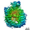 10688MC  6y53C 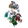 6y5qC M: map data used to model this data C: citing same article ( |
|---|---|
| Similar structure data |

-Links
 PDBj
PDBj


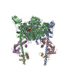
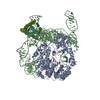
- Assembly
| Deposited unit | 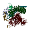
|
|---|---|
| 1 |
|
-Components
-Protein , 4 types, 4 molecules 9yqp
| #1: Protein | Mass: 58934.844 Da / Num. of mol.: 1 / Source method: isolated from a natural source / Details: SF3A3, SAP61 / Source: (natural) Homo sapiens (human) / References: UniProt: Q12874 |
|---|---|
| #2: Protein | Mass: 12427.524 Da / Num. of mol.: 1 / Source method: isolated from a natural source / Details: PHF5A / Source: (natural) Homo sapiens (human) / References: UniProt: Q7RTV0 |
| #8: Protein | Mass: 85965.961 Da / Num. of mol.: 1 / Source method: isolated from a natural source / Details: HTATSF1 / Source: (natural) Homo sapiens (human) / References: UniProt: O43719 |
| #9: Protein | Mass: 117576.484 Da / Num. of mol.: 1 / Source method: isolated from a natural source / Details: DDX46, KIAA0801 / Source: (natural) Homo sapiens (human) / References: UniProt: Q7L014, RNA helicase |
-Splicing factor 3B subunit ... , 4 types, 4 molecules x8vu
| #3: Protein | Mass: 10149.369 Da / Num. of mol.: 1 / Source method: isolated from a natural source / Details: SF3B5, SF3B10 / Source: (natural) Homo sapiens (human) / References: UniProt: Q9BWJ5 |
|---|---|
| #4: Protein | Mass: 100377.812 Da / Num. of mol.: 1 / Source method: isolated from a natural source / Details: SF3B2, SAP145 / Source: (natural) Homo sapiens (human) / References: UniProt: Q13435 |
| #5: Protein | Mass: 135718.844 Da / Num. of mol.: 1 / Source method: isolated from a natural source / Details: SF3B3, KIAA0017, SAP130 / Source: (natural) Homo sapiens (human) / References: UniProt: Q15393 |
| #6: Protein | Mass: 146024.938 Da / Num. of mol.: 1 / Source method: isolated from a natural source / Details: SF3B1, SAP155 / Source: (natural) Homo sapiens (human) / References: UniProt: O75533 |
-RNA chain / Non-polymers , 2 types, 4 molecules 2
| #10: Chemical |  Mass: 65.409 Da / Num. of mol.: 3 / Source method: isolated from a natural source / Formula: Zn Mass: 65.409 Da / Num. of mol.: 3 / Source method: isolated from a natural source / Formula: Zn#7: RNA chain | | Mass: 60186.445 Da / Num. of mol.: 1 / Source method: isolated from a natural source / Source: (natural) Homo sapiens (human) / References: GenBank: 36516 |
|---|
-Details
| Has ligand of interest | N |
|---|
-Experimental details
-Experiment
| Experiment | Method: ELECTRON MICROSCOPY |
|---|---|
| EM experiment | Aggregation state: PARTICLE / 3D reconstruction method: single particle reconstruction |
- Sample preparation
| Component | Name: human 17S U2 snRNP / Type: COMPLEX / Entity ID: #1-#9 / Source: NATURAL |
|---|---|
| Source (natural) | Organism: Homo sapiens (human) |
| Buffer solution | pH: 7.9 |
| Specimen | Embedding applied: NO / Shadowing applied: NO / Staining applied: NO / Vitrification applied: YES |
| Specimen support | Grid material: COPPER / Grid type: Quantifoil R3.5/1 |
| Vitrification | Instrument: FEI VITROBOT MARK I / Cryogen name: ETHANE / Humidity: 100 % / Chamber temperature: 277 K |
- Electron microscopy imaging
| Experimental equipment |  Model: Titan Krios / Image courtesy: FEI Company |
|---|---|
| Microscopy | Model: FEI TITAN KRIOS |
| Electron gun | Electron source:  FIELD EMISSION GUN / Accelerating voltage: 300 kV / Illumination mode: SPOT SCAN FIELD EMISSION GUN / Accelerating voltage: 300 kV / Illumination mode: SPOT SCAN |
| Electron lens | Mode: BRIGHT FIELD / Cs: 0.01 mm / Alignment procedure: ZEMLIN TABLEAU |
| Specimen holder | Cryogen: NITROGEN / Specimen holder model: FEI TITAN KRIOS AUTOGRID HOLDER |
| Image recording | Average exposure time: 1 sec. / Electron dose: 72 e/Å2 / Detector mode: INTEGRATING / Film or detector model: FEI FALCON III (4k x 4k) |
- Processing
| EM software |
| ||||||||||||||||||||
|---|---|---|---|---|---|---|---|---|---|---|---|---|---|---|---|---|---|---|---|---|---|
| CTF correction | Type: PHASE FLIPPING AND AMPLITUDE CORRECTION | ||||||||||||||||||||
| Symmetry | Point symmetry: C1 (asymmetric) | ||||||||||||||||||||
| 3D reconstruction | Resolution: 4.1 Å / Resolution method: FSC 0.143 CUT-OFF / Num. of particles: 120070 / Symmetry type: POINT | ||||||||||||||||||||
| Atomic model building | Space: REAL |
