2YNP
 
 | | yeast betaprime COP 1-604 with KTKTN motif | | 分子名称: | COATOMER SUBUNIT BETA', KTKTN MOTIF | | 著者 | Jackson, L.P, Lewis, M, Kent, H.M, Edeling, M.A, Evans, P.R, Duden, R, Owen, D.J. | | 登録日 | 2012-10-17 | | 公開日 | 2012-12-12 | | 最終更新日 | 2023-12-20 | | 実験手法 | X-RAY DIFFRACTION (2.962 Å) | | 主引用文献 | Molecular Basis for Recognition of Dilysine Trafficking Motifs by Copi.
Dev.Cell, 23, 2012
|
|
6X08
 | |
2XU7
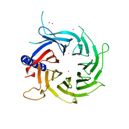
 | | Structural basis for RbAp48 binding to FOG-1 | | 分子名称: | HISTONE-BINDING PROTEIN RBBP4, TETRAETHYLENE GLYCOL, ZINC FINGER PROTEIN ZFPM1 | | 著者 | Lejon, S, Thong, S.Y, Murthy, A, Blobel, G.A, Mackay, J.P, Murzina, N.V, Laue, E.D. | | 登録日 | 2010-10-15 | | 公開日 | 2010-11-03 | | 最終更新日 | 2023-12-20 | | 実験手法 | X-RAY DIFFRACTION (1.9 Å) | | 主引用文献 | Insights Into Association of the Nurd Complex with Fog-1 from the Crystal Structure of an Rbap48-Fog- 1 Complex.
J.Biol.Chem., 286, 2011
|
|
2XYI
 | | Crystal Structure of Nurf55 in complex with a H4 peptide | | 分子名称: | DI(HYDROXYETHYL)ETHER, HISTONE H4, PROBABLE HISTONE-BINDING PROTEIN CAF1, ... | | 著者 | Stirnimann, C.U, Nowak, A.J, Mueller, C.W. | | 登録日 | 2010-11-17 | | 公開日 | 2011-05-04 | | 最終更新日 | 2023-12-20 | | 実験手法 | X-RAY DIFFRACTION (1.75 Å) | | 主引用文献 | Chromatin-Modifying Complex Component Nurf55/P55 Associates with Histones H3, H4 and Polycomb Repressive Complex 2 Subunit Su(Z)12 Through Partially Overlapping Binding Sites.
J.Biol.Chem., 286, 2011
|
|
2YBA
 | | Crystal structure of Nurf55 in complex with histone H3 | | 分子名称: | HISTONE H3, PROBABLE HISTONE-BINDING PROTEIN CAF1 | | 著者 | Schmitges, F.W, Prusty, A.B, Faty, M, Stutzer, A, Lingaraju, G.M, Aiwazian, J, Sack, R, Hess, D, Li, L, Zhou, S, Bunker, R.D, Wirth, U, Bouwmeester, T, Bauer, A, Ly-Hartig, N, Zhao, K, Chan, H, Gu, J, Gut, H, Fischle, W, Muller, J, Thoma, N.H. | | 登録日 | 2011-03-02 | | 公開日 | 2011-05-11 | | 最終更新日 | 2024-05-01 | | 実験手法 | X-RAY DIFFRACTION (2.55 Å) | | 主引用文献 | Histone Methylation by Prc2 is Inhibited by Active Chromatin Marks
Mol.Cell, 42, 2011
|
|
2OVQ
 | | Structure of the Skp1-Fbw7-CyclinEdegC complex | | 分子名称: | F-box/WD repeat protein 7, S-phase kinase-associated protein 1A, SULFATE ION, ... | | 著者 | Hao, B, Oehlmann, S, Sowa, M.E, Harper, J.W, Pavletich, N.P. | | 登録日 | 2007-02-14 | | 公開日 | 2007-04-24 | | 最終更新日 | 2024-04-03 | | 実験手法 | X-RAY DIFFRACTION (2.6 Å) | | 主引用文献 | Structure of a Fbw7-Skp1-Cyclin E Complex: Multisite-Phosphorylated Substrate Recognition by SCF Ubiquitin Ligases
Mol.Cell, 26, 2007
|
|
2OVP
 | | Structure of the Skp1-Fbw7 complex | | 分子名称: | F-box/WD repeat protein 7, S-phase kinase-associated protein 1A | | 著者 | Hao, B, Oehlmann, S, Sowa, M.E, Harper, J.W, Pavletich, N.P. | | 登録日 | 2007-02-14 | | 公開日 | 2007-04-24 | | 最終更新日 | 2024-04-03 | | 実験手法 | X-RAY DIFFRACTION (2.9 Å) | | 主引用文献 | Structure of a Fbw7-Skp1-Cyclin E Complex: Multisite-Phosphorylated Substrate Recognition by SCF Ubiquitin Ligases
Mol.Cell, 26, 2007
|
|
2PBI
 | | The multifunctional nature of Gbeta5/RGS9 revealed from its crystal structure | | 分子名称: | GLYCEROL, Guanine nucleotide-binding protein subunit beta 5, Regulator of G-protein signaling 9 | | 著者 | Cheever, M.L, Snyder, J.T, Gershburg, S, Siderovski, D.P, Harden, T.K, Sondek, J. | | 登録日 | 2007-03-28 | | 公開日 | 2008-01-29 | | 最終更新日 | 2024-02-21 | | 実験手法 | X-RAY DIFFRACTION (1.95 Å) | | 主引用文献 | Crystal structure of the multifunctional Gbeta5-RGS9 complex.
Nat.Struct.Mol.Biol., 15, 2008
|
|
3BG1
 | |
3BG0
 | |
2OVR
 | | Structure of the Skp1-Fbw7-CyclinEdegN complex | | 分子名称: | F-box/WD repeat protein 7, S-phase kinase-associated protein 1A, SULFATE ION, ... | | 著者 | Hao, B, Oehlmann, S, Sowa, M.E, Harper, J.W, Pavletich, N.P. | | 登録日 | 2007-02-14 | | 公開日 | 2007-04-24 | | 最終更新日 | 2024-04-03 | | 実験手法 | X-RAY DIFFRACTION (2.5 Å) | | 主引用文献 | Structure of a Fbw7-Skp1-Cyclin E Complex: Multisite-Phosphorylated Substrate Recognition by SCF Ubiquitin Ligases
Mol.Cell, 26, 2007
|
|
3AH8
 | | Structure of heterotrimeric G protein Galpha-q beta gamma in complex with an inhibitor YM-254890 | | 分子名称: | GUANOSINE-5'-DIPHOSPHATE, Guanine nucleotide-binding protein G(I)/G(S)/G(O) subunit gamma-2, Guanine nucleotide-binding protein G(I)/G(S)/G(T) subunit beta-1, ... | | 著者 | Nishimura, A, Kitano, K, Takasaki, J, Taniguchi, M, Mizuno, N, Tago, K, Hakoshima, T, Itoh, H. | | 登録日 | 2010-04-20 | | 公開日 | 2010-07-21 | | 最終更新日 | 2023-11-15 | | 実験手法 | X-RAY DIFFRACTION (2.9 Å) | | 主引用文献 | Structural basis for the specific inhibition of heterotrimeric Gq protein by a small molecule.
Proc.Natl.Acad.Sci.USA, 107, 2010
|
|
3C99
 | |
2PM7
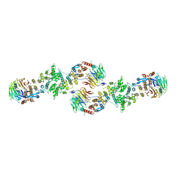
 | | Crystal structure of yeast Sec13/31 edge element of the COPII vesicular coat, selenomethionine version | | 分子名称: | Protein transport protein SEC13, Protein transport protein SEC31 | | 著者 | Goldberg, J, Fath, S, Mancias, J.D, Bi, X. | | 登録日 | 2007-04-20 | | 公開日 | 2007-07-03 | | 最終更新日 | 2021-10-20 | | 実験手法 | X-RAY DIFFRACTION (2.35 Å) | | 主引用文献 | Structure and Organization of Coat Proteins in the COPII Cage.
Cell(Cambridge,Mass.), 129, 2007
|
|
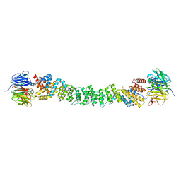
2PM6
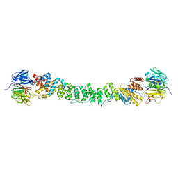 | | Crystal Structure of yeast Sec13/31 edge element of the COPII vesicular coat, native version | | 分子名称: | Protein transport protein SEC13, Protein transport protein SEC31 | | 著者 | Goldberg, J, Fath, S, Mancias, J.D, Bi, X. | | 登録日 | 2007-04-20 | | 公開日 | 2007-07-03 | | 最終更新日 | 2024-04-03 | | 実験手法 | X-RAY DIFFRACTION (2.45 Å) | | 主引用文献 | Structure and Organization of Coat Proteins in the COPII Cage.
Cell(Cambridge,Mass.), 129, 2007
|
|
2I3T
 | |
2I3S
 | | Bub3 complex with Bub1 GLEBS motif | | 分子名称: | Cell cycle arrest protein, Checkpoint serine/threonine-protein kinase | | 著者 | Larsen, N.A, Harrison, S.C. | | 登録日 | 2006-08-20 | | 公開日 | 2007-01-09 | | 最終更新日 | 2023-08-30 | | 実験手法 | X-RAY DIFFRACTION (1.9 Å) | | 主引用文献 | Structural analysis of Bub3 interactions in the mitotic spindle checkpoint.
Proc.Natl.Acad.Sci.Usa, 104, 2007
|
|
4JSP
 | |
4JT5
 | | mTORdeltaN-mLST8-pp242 complex | | 分子名称: | 2-[4-amino-1-(propan-2-yl)-1H-pyrazolo[3,4-d]pyrimidin-3-yl]-1H-indol-5-ol, Serine/threonine-protein kinase mTOR, Target of rapamycin complex subunit LST8 | | 著者 | Pavletich, N.P, Yang, H. | | 登録日 | 2013-03-22 | | 公開日 | 2013-05-08 | | 最終更新日 | 2024-02-28 | | 実験手法 | X-RAY DIFFRACTION (3.45 Å) | | 主引用文献 | mTOR kinase structure, mechanism and regulation.
Nature, 497, 2013
|
|
4L9O
 | | Crystal Structure of the Sec13-Sec16 blade-inserted complex from Pichia pastoris | | 分子名称: | 1,2-ETHANEDIOL, CALCIUM ION, CHLORIDE ION, ... | | 著者 | McMahon, C, Jeffrey, P.D, Hughson, F.M. | | 登録日 | 2013-06-18 | | 公開日 | 2013-10-02 | | 最終更新日 | 2023-09-20 | | 実験手法 | X-RAY DIFFRACTION (1.6 Å) | | 主引用文献 | Sec16 influences transitional ER sites by regulating rather than organizing COPII.
Mol Biol Cell, 24, 2013
|
|
4JSX
 | | structure of mTORDeltaN-mLST8-Torin2 complex | | 分子名称: | 9-(6-aminopyridin-3-yl)-1-[3-(trifluoromethyl)phenyl]benzo[h][1,6]naphthyridin-2(1H)-one, Serine/threonine-protein kinase mTOR, Target of rapamycin complex subunit LST8 | | 著者 | Pavletich, N.P, Yang, H. | | 登録日 | 2013-03-22 | | 公開日 | 2013-05-08 | | 最終更新日 | 2024-02-28 | | 実験手法 | X-RAY DIFFRACTION (3.5 Å) | | 主引用文献 | mTOR kinase structure, mechanism and regulation.
Nature, 497, 2013
|
|
4N14
 | | Crystal structure of Cdc20 and apcin complex | | 分子名称: | 2-(2-methyl-5-nitro-1H-imidazol-1-yl)ethyl [(1R)-2,2,2-trichloro-1-(pyrimidin-2-ylamino)ethyl]carbamate, Cell division cycle protein 20 homolog | | 著者 | Luo, X, Tian, W, Yu, H. | | 登録日 | 2013-10-03 | | 公開日 | 2014-08-20 | | 最終更新日 | 2024-02-28 | | 実験手法 | X-RAY DIFFRACTION (2.1 Å) | | 主引用文献 | Synergistic blockade of mitotic exit by two chemical inhibitors of the APC/C.
Nature, 514, 2014
|
|
4KFM
 | |
3W15
 | | Structure of peroxisomal targeting signal 2 (PTS2) of Saccharomyces cerevisiae 3-ketoacyl-CoA thiolase in complex with Pex7p and Pex21p | | 分子名称: | 3-ketoacyl-CoA thiolase, peroxisomal, Maltose-binding periplasmic protein, ... | | 著者 | Pan, D, Nakatsu, T, Kato, H. | | 登録日 | 2012-11-06 | | 公開日 | 2013-07-03 | | 最終更新日 | 2017-08-16 | | 実験手法 | X-RAY DIFFRACTION (1.8 Å) | | 主引用文献 | Crystal structure of peroxisomal targeting signal-2 bound to its receptor complex Pex7p-Pex21p
Nat.Struct.Mol.Biol., 20, 2013
|
|
3V7D
 | | Crystal Structure of ScSkp1-ScCdc4-pSic1 peptide complex | | 分子名称: | Cell division control protein 4, Protein SIC1, Suppressor of kinetochore protein 1 | | 著者 | Tang, X, Orlicky, S, Mittag, T, Csizmok, V, Pawson, T, Forman-Kay, J, Sicheri, F, Tyers, M. | | 登録日 | 2011-12-20 | | 公開日 | 2012-05-02 | | 最終更新日 | 2023-09-13 | | 実験手法 | X-RAY DIFFRACTION (2.306 Å) | | 主引用文献 | Composite low affinity interactions dictate recognition of the cyclin-dependent kinase inhibitor Sic1 by the SCFCdc4 ubiquitin ligase.
Proc.Natl.Acad.Sci.USA, 109, 2012
|
|


