 ムービー
ムービー コントローラー
 構造ビューア
 EMN検索について


-検索条件
-検索結果
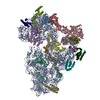
検索 (著者・登録者: sander & b)の結果514件中、1から50件目までを表示しています

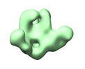
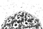
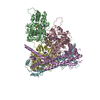
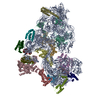
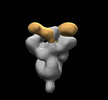
EMDB-42464: 
chEnv TTT protein in complex with 43A2 Fab

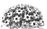
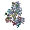
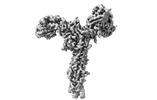
EMDB-42468:
chEnv TTT protein in complex with CM01A Fab
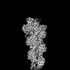
EMDB-18482:
Herpes simplex virus 1 capsid (WT) vertices in perinuclear NEC-coated vesicles determined in situ

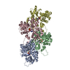
EMDB-18484:
Herpes simplex virus 1 nuclear egress complex (WT) determined in situ from perinuclear vesicles
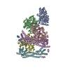
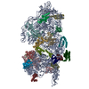
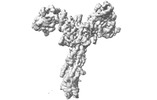
EMDB-43664:
Triple tandem trimer immunogens for HIV-1 and influenza nucleic acid-based vaccines

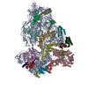
EMDB-43665:
Triple tandem trimer immunogens for HIV-1 and influenza nucleic acid-based vaccines (cH125 TTT)
EMDB-43666:
Triple tandem trimer immunogens for HIV-1 and influenza nucleic acid-based vaccines (H2/1 GCN4)
EMDB-43668:
Triple tandem trimer immunogens for HIV-1 and influenza nucleic acid-based vaccines (H5/1 GCN4)
EMDB-43669:
Triple tandem trimer immunogens for HIV-1 and influenza nucleic acid-based vaccines. H5 GCN4
EMDB-17974:
Pseudorabies virus cytosolic C-capsid (US3 KO) vertices determined in situ

EMDB-17975:
Pseudorabies virus primary enveloped (perinuclear) C-capsid (US3 KO) vertices determined in situ
EMDB-17976:
Pseudorabies nuclear C-capsids (US3 KO) vertices determined in situ

EMDB-18473:
Subtomogram average of pseudorabies virus nuclear egress complex helical form (UL31/34) determined in situ

EMDB-18474:
Subtomogram average of pseudorabies virus nuclear egress complex (UL31/34) determined in situ

EMDB-18479:
Pseudorabies virus cytosolic C-capsid (WT) vertices determined in situ
EMDB-18480:
Pseudorabies virus nuclear C-capsid (WT) vertices determined in situ
EMDB-18481:
Herpes simplex virus 1 cytosolic C-capsid (WT) vertices determined in situ
EMDB-18483:
Herpes simplex virus 1 nuclear C-capsid (WT) vertices determined in situ
EMDB-19497:
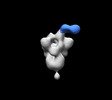
Cryo-EM reconstruction of the formin Cdc12 bound to the barbed end of F-actin (without phalloidin)

EMDB-19499:
Structure of the F-actin barbed end bound by Cdc12 and profilin (ring complex) at a resolution of 6.3 Angstrom

EMDB-19501:
Structure of the undecorated barbed end of F-actin.

EMDB-19503:
Structure of the F-actin barbed end bound by formin mDia1

EMDB-19522:
Structure of the formin INF2 bound to the barbed end of F-actin.

PDB-8rty:
Structure of the F-actin barbed end bound by Cdc12 and profilin (ring complex) at a resolution of 6.3 Angstrom
PDB-8ru0:
Structure of the undecorated barbed end of F-actin.
PDB-8ru2:
Structure of the F-actin barbed end bound by formin mDia1
PDB-8rv2:
Structure of the formin INF2 bound to the barbed end of F-actin.

EMDB-19496:
Structure of the formin Cdc12 bound to the barbed end of phalloidin-stabilized F-actin.
PDB-8rtt:
Structure of the formin Cdc12 bound to the barbed end of phalloidin-stabilized F-actin.

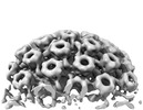
PDB-8qox:
Two-component assembly of SlaA and SlaB S-layer proteins of Sulfolobus acidocaldarius

PDB-8qp0:
A hexamer pore in the S-layer of Sulfolobus acidocaldarius formed by SlaA protein
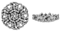
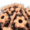
EMDB-18127:
S-layer of archaeon Sulfolobus acidocaldarius by subtomogram averaging

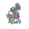
EMDB-16595:
Rnase R bound to a 30S degradation intermediate (main state)
EMDB-16596:
Rnase R bound to a 30S degradation intermediate (state II)
EMDB-16605:
Rnase R bound to a 30S degradation intermediate (State I - head-turning)
EMDB-16606:
Rnase R bound to a 30S degradation intermediate (State I - head-turning)
EMDB-16607:
Rnase R bound to a 30S degradation intermediate (State I - head-turning)
PDB-8cdu:
Rnase R bound to a 30S degradation intermediate (main state)
PDB-8cdv:
Rnase R bound to a 30S degradation intermediate (state II)
PDB-8cec:
Rnase R bound to a 30S degradation intermediate (State I - head-turning)
PDB-8ced:
Rnase R bound to a 30S degradation intermediate (State I - head-turning)
PDB-8cee:
Rnase R bound to a 30S degradation intermediate (State I - head-turning)
EMDB-28850:
SARS-CoV-2 Gamma 6P Mut7 S + COVA309-3 Fab
EMDB-28851:
SARS-CoV-2 Gamma 6P Mut7 S + COVA309-10 Fab
EMDB-28852:
SARS-CoV-2 Omicron 6P S + COVA309-35 Fab
EMDB-28853:
SARS-CoV-2 Gamma 6P Mut7 + S COVA309-38 Fab
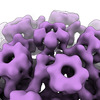
EMDB-15530:
S-layer protein SlaA from Sulfolobus acidocaldarius at pH 10.0
EMDB-15531:
S-layer protein SlaA from Sulfolobus acidocaldarius at pH 7.0

PDB-8an2:
S-layer protein SlaA from Sulfolobus acidocaldarius at pH 10.0

PDB-8an3:
S-layer protein SlaA from Sulfolobus acidocaldarius at pH 7.0
ページ:
 wwPDBはEMDBデータモデルのバージョン3へ移行します
