Movie
Movie Controller
 Structure viewers
 About Yorodumi Papers


+Search query
-Structure paper
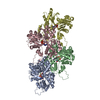
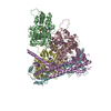
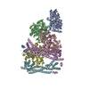
| Title | Molecular mechanism of actin filament elongation by formins. |
|---|---|
| Journal, issue, pages | Science, Vol. 384, Issue 6692, Page eadn9560, Year 2024 |
| Publish date | Apr 12, 2024 |
 Authors Authors | Wout Oosterheert / Micaela Boiero Sanders / Johanna Funk / Daniel Prumbaum / Stefan Raunser / Peter Bieling /  |
| PubMed Abstract | Formins control the assembly of actin filaments (F-actin) that drive cell morphogenesis and motility in eukaryotes. However, their molecular interaction with F-actin and their mechanism of action ...Formins control the assembly of actin filaments (F-actin) that drive cell morphogenesis and motility in eukaryotes. However, their molecular interaction with F-actin and their mechanism of action remain unclear. In this work, we present high-resolution cryo-electron microscopy structures of F-actin barbed ends bound by three distinct formins, revealing a common asymmetric formin conformation imposed by the filament. Formation of new intersubunit contacts during actin polymerization sterically displaces formin and triggers its translocation. This "undock-and-lock" mechanism explains how actin-filament growth is coordinated with formin movement. Filament elongation speeds are controlled by the positioning and stability of actin-formin interfaces, which distinguish fast and slow formins. Furthermore, we provide a structure of the actin-formin-profilin ring complex, which resolves how profilin is rapidly released from the barbed end during filament elongation. |
 External links External links | Science / PubMed:38603491 |
| Methods | EM (single particle) |
| Resolution | 3.08 - 6.25 Å |
| Structure data |  EMDB-19497: Cryo-EM reconstruction of the formin Cdc12 bound to the barbed end of F-actin (without phalloidin) EMDB-19499, PDB-8rty: EMDB-19501, PDB-8ru0: EMDB-19503, PDB-8ru2: EMDB-19522, PDB-8rv2: |
| Chemicals |  ChemComp-ADP:  ChemComp-MG:  ChemComp-PO4:  ChemComp-ATP: |
| Source |
|
 Keywords Keywords |  STRUCTURAL PROTEIN / actin / formin / Cdc12 / profilin / actin assembly / actin end / barbed end / INF2 STRUCTURAL PROTEIN / actin / formin / Cdc12 / profilin / actin assembly / actin end / barbed end / INF2 |