Movie
Movie Controller

[English] 日本語
 Yorodumi
- EMDB-19497: Cryo-EM reconstruction of the formin Cdc12 bound to the barbed en... -
+ Open data

- Basic information
| Entry | 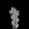 | ||||||||||||
|---|---|---|---|---|---|---|---|---|---|---|---|---|---|
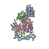
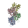
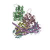
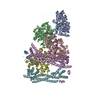
| Title | Cryo-EM reconstruction of the formin Cdc12 bound to the barbed end of F-actin (without phalloidin) | ||||||||||||
 Map data Map data | Sharpened cryo-EM density map of the formin Cdc12 bound to the barbed end of F-actin (without phalloidin). | ||||||||||||
 Sample Sample |
| ||||||||||||
 Keywords Keywords |  actin / formin / Cdc12 / actin assembly. / STRUCTURAL PROTEIN actin / formin / Cdc12 / actin assembly. / STRUCTURAL PROTEIN | ||||||||||||
| Function / homology |  Function and homology informationF-bar domain binding / protein localization to mitotic actomyosin contractile ring / medial cortical node / mitotic actomyosin contractile ring, proximal layer / mitotic actomyosin contractile ring / medial cortex / mitotic actomyosin contractile ring assembly / positive regulation of norepinephrine uptake / cellular response to cytochalasin B / bBAF complex ...F-bar domain binding / protein localization to mitotic actomyosin contractile ring / medial cortical node / mitotic actomyosin contractile ring, proximal layer / mitotic actomyosin contractile ring / medial cortex / mitotic actomyosin contractile ring assembly / positive regulation of norepinephrine uptake / cellular response to cytochalasin B / bBAF complex / npBAF complex / postsynaptic actin cytoskeleton organization / regulation of transepithelial transport / brahma complex / nBAF complex / structural constituent of postsynaptic actin cytoskeleton / morphogenesis of a polarized epithelium / Formation of annular gap junctions / GBAF complex / Gap junction degradation / postsynaptic actin cytoskeleton / protein localization to adherens junction / regulation of G0 to G1 transition / dense body / Cell-extracellular matrix interactions / Tat protein binding / Folding of actin by CCT/TriC / regulation of double-strand break repair / regulation of nucleotide-excision repair / RSC-type complex / apical protein localization / mating projection tip / Prefoldin mediated transfer of substrate to CCT/TriC / barbed-end actin filament capping / adherens junction assembly / RHOF GTPase cycle / Adherens junctions interactions / tight junction / Sensory processing of sound by outer hair cells of the cochlea / SWI/SNF complex / Interaction between L1 and Ankyrins / Sensory processing of sound by inner hair cells of the cochlea / regulation of mitotic metaphase/anaphase transition / regulation of norepinephrine uptake / positive regulation of double-strand break repair / positive regulation of T cell differentiation / NuA4 histone acetyltransferase complex / regulation of synaptic vesicle endocytosis / apical junction complex / maintenance of blood-brain barrier / establishment or maintenance of cell polarity / cell division site / cortical cytoskeleton / positive regulation of double-strand break repair via homologous recombination / positive regulation of stem cell population maintenance / nitric-oxide synthase binding / Recycling pathway of L1 / regulation of cyclin-dependent protein serine/threonine kinase activity / regulation of G1/S transition of mitotic cell cycle / actin filament bundle assembly / negative regulation of cell differentiation / brush border / kinesin binding / calyx of Held / EPH-ephrin mediated repulsion of cells / RHO GTPases Activate WASPs and WAVEs / RHO GTPases activate IQGAPs / positive regulation of myoblast differentiation / regulation of protein localization to plasma membrane / EPHB-mediated forward signaling / substantia nigra development / actin filament polymerization / axonogenesis / negative regulation of protein binding / actin filament / cell motility / RHO GTPases Activate Formins / Translocation of SLC2A4 (GLUT4) to the plasma membrane / regulation of transmembrane transporter activity / positive regulation of cell differentiation / FCGR3A-mediated phagocytosis / adherens junction / Hydrolases; Acting on acid anhydrides; Acting on acid anhydrides to facilitate cellular and subcellular movement / DNA Damage Recognition in GG-NER / tau protein binding / Signaling by high-kinase activity BRAF mutants / Schaffer collateral - CA1 synapse / MAP2K and MAPK activation / B-WICH complex positively regulates rRNA expression / structural constituent of cytoskeleton / cytoplasmic ribonucleoprotein granule / kinetochore / Regulation of actin dynamics for phagocytic cup formation / platelet aggregation / small GTPase binding / nuclear matrix / VEGFA-VEGFR2 Pathway / UCH proteinases / Signaling by RAF1 mutants / Signaling by moderate kinase activity BRAF mutants Function and homology informationF-bar domain binding / protein localization to mitotic actomyosin contractile ring / medial cortical node / mitotic actomyosin contractile ring, proximal layer / mitotic actomyosin contractile ring / medial cortex / mitotic actomyosin contractile ring assembly / positive regulation of norepinephrine uptake / cellular response to cytochalasin B / bBAF complex ...F-bar domain binding / protein localization to mitotic actomyosin contractile ring / medial cortical node / mitotic actomyosin contractile ring, proximal layer / mitotic actomyosin contractile ring / medial cortex / mitotic actomyosin contractile ring assembly / positive regulation of norepinephrine uptake / cellular response to cytochalasin B / bBAF complex / npBAF complex / postsynaptic actin cytoskeleton organization / regulation of transepithelial transport / brahma complex / nBAF complex / structural constituent of postsynaptic actin cytoskeleton / morphogenesis of a polarized epithelium / Formation of annular gap junctions / GBAF complex / Gap junction degradation / postsynaptic actin cytoskeleton / protein localization to adherens junction / regulation of G0 to G1 transition / dense body / Cell-extracellular matrix interactions / Tat protein binding / Folding of actin by CCT/TriC / regulation of double-strand break repair / regulation of nucleotide-excision repair / RSC-type complex / apical protein localization / mating projection tip / Prefoldin mediated transfer of substrate to CCT/TriC / barbed-end actin filament capping / adherens junction assembly / RHOF GTPase cycle / Adherens junctions interactions / tight junction / Sensory processing of sound by outer hair cells of the cochlea / SWI/SNF complex / Interaction between L1 and Ankyrins / Sensory processing of sound by inner hair cells of the cochlea / regulation of mitotic metaphase/anaphase transition / regulation of norepinephrine uptake / positive regulation of double-strand break repair / positive regulation of T cell differentiation / NuA4 histone acetyltransferase complex / regulation of synaptic vesicle endocytosis / apical junction complex / maintenance of blood-brain barrier / establishment or maintenance of cell polarity / cell division site / cortical cytoskeleton / positive regulation of double-strand break repair via homologous recombination / positive regulation of stem cell population maintenance / nitric-oxide synthase binding / Recycling pathway of L1 / regulation of cyclin-dependent protein serine/threonine kinase activity / regulation of G1/S transition of mitotic cell cycle / actin filament bundle assembly / negative regulation of cell differentiation / brush border / kinesin binding / calyx of Held / EPH-ephrin mediated repulsion of cells / RHO GTPases Activate WASPs and WAVEs / RHO GTPases activate IQGAPs / positive regulation of myoblast differentiation / regulation of protein localization to plasma membrane / EPHB-mediated forward signaling / substantia nigra development / actin filament polymerization / axonogenesis / negative regulation of protein binding / actin filament / cell motility / RHO GTPases Activate Formins / Translocation of SLC2A4 (GLUT4) to the plasma membrane / regulation of transmembrane transporter activity / positive regulation of cell differentiation / FCGR3A-mediated phagocytosis / adherens junction / Hydrolases; Acting on acid anhydrides; Acting on acid anhydrides to facilitate cellular and subcellular movement / DNA Damage Recognition in GG-NER / tau protein binding / Signaling by high-kinase activity BRAF mutants / Schaffer collateral - CA1 synapse / MAP2K and MAPK activation / B-WICH complex positively regulates rRNA expression / structural constituent of cytoskeleton / cytoplasmic ribonucleoprotein granule / kinetochore / Regulation of actin dynamics for phagocytic cup formation / platelet aggregation / small GTPase binding / nuclear matrix / VEGFA-VEGFR2 Pathway / UCH proteinases / Signaling by RAF1 mutants / Signaling by moderate kinase activity BRAF mutantsSimilarity search - Function | ||||||||||||
| Biological species |  Homo sapiens (human) / Homo sapiens (human) /  Schizosaccharomyces pombe (fission yeast) Schizosaccharomyces pombe (fission yeast) | ||||||||||||
| Method | single particle reconstruction / cryo EM / Resolution: 4.54 Å | ||||||||||||
 Authors Authors | Oosterheert W / Boiero Sanders M / Funk J / Prumbaum D / Raunser S / Bieling P | ||||||||||||
| Funding support |  Germany, European Union, 3 items Germany, European Union, 3 items
| ||||||||||||
 Citation Citation | Journal: Science / Year: 2024 Title: Molecular mechanism of actin filament elongation by formins. Authors: Wout Oosterheert / Micaela Boiero Sanders / Johanna Funk / Daniel Prumbaum / Stefan Raunser / Peter Bieling / Abstract: Formins control the assembly of actin filaments (F-actin) that drive cell morphogenesis and motility in eukaryotes. However, their molecular interaction with F-actin and their mechanism of action ...Formins control the assembly of actin filaments (F-actin) that drive cell morphogenesis and motility in eukaryotes. However, their molecular interaction with F-actin and their mechanism of action remain unclear. In this work, we present high-resolution cryo-electron microscopy structures of F-actin barbed ends bound by three distinct formins, revealing a common asymmetric formin conformation imposed by the filament. Formation of new intersubunit contacts during actin polymerization sterically displaces formin and triggers its translocation. This "undock-and-lock" mechanism explains how actin-filament growth is coordinated with formin movement. Filament elongation speeds are controlled by the positioning and stability of actin-formin interfaces, which distinguish fast and slow formins. Furthermore, we provide a structure of the actin-formin-profilin ring complex, which resolves how profilin is rapidly released from the barbed end during filament elongation. | ||||||||||||
| History |
|
- Structure visualization
| Supplemental images |
|---|

- Downloads & links
-EMDB archive
| Map data | emd_19497.map.gz | 13.3 MB | EMDB map data format | |
|---|---|---|---|---|
| Header (meta data) | emd-19497-v30.xmlemd-19497.xml | 31.1 KB 31.1 KB | Display Display | EMDB header |
| FSC (resolution estimation) | emd_19497_fsc.xml | 11.5 KB | Display | FSC data file |
| Images | 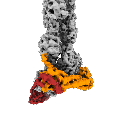 emd_19497.png emd_19497.png | 82.9 KB | ||
| Masks | emd_19497_msk_1.map | 125 MB | Mask map | |
| Filedesc metadata | emd-19497.cif.gz | 7 KB | ||
| Others | emd_19497_additional_1.map.gzemd_19497_additional_2.map.gzemd_19497_additional_3.map.gzemd_19497_additional_4.map.gzemd_19497_half_map_1.map.gzemd_19497_half_map_2.map.gz | 97.3 MB 107.5 MB 113.7 MB 115.5 MB 98.5 MB 98.5 MB | ||
| Archive directory |  http://ftp.pdbj.org/pub/emdb/structures/EMD-19497ftp://ftp.pdbj.org/pub/emdb/structures/EMD-19497 http://ftp.pdbj.org/pub/emdb/structures/EMD-19497ftp://ftp.pdbj.org/pub/emdb/structures/EMD-19497 | HTTPS FTP |
-Related structure data


-Links
| EMDB pages | EMDB (EBI/PDBe) / EMDataResource |
|---|---|
| Related items in Molecule of the Month |


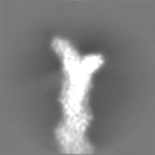
-Map
| File | Download / File: emd_19497.map.gz / Format: CCP4 / Size: 125 MB / Type: IMAGE STORED AS FLOATING POINT NUMBER (4 BYTES) | ||||||||||||||||||||||||||||||||||||
|---|---|---|---|---|---|---|---|---|---|---|---|---|---|---|---|---|---|---|---|---|---|---|---|---|---|---|---|---|---|---|---|---|---|---|---|---|---|
| Annotation | Sharpened cryo-EM density map of the formin Cdc12 bound to the barbed end of F-actin (without phalloidin). | ||||||||||||||||||||||||||||||||||||
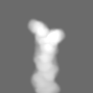
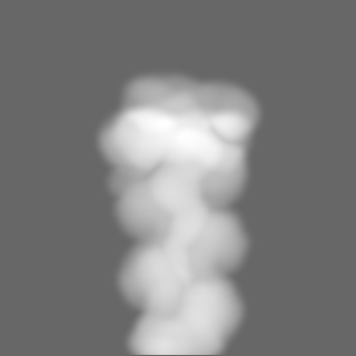
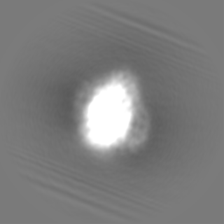
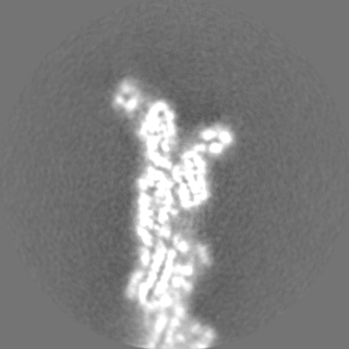
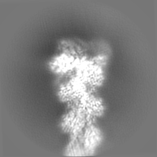
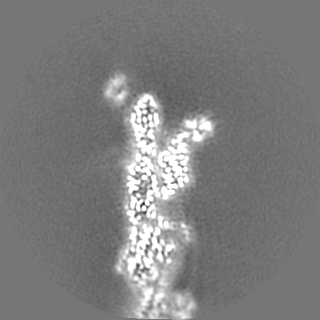
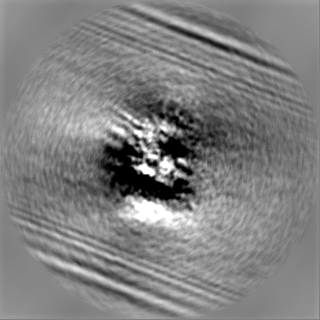
| Projections & slices | Image control
Images are generated by Spider. | ||||||||||||||||||||||||||||||||||||
| Voxel size | X=Y=Z: 0.88 Å | ||||||||||||||||||||||||||||||||||||
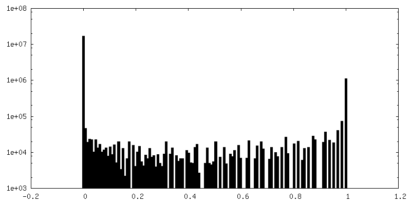
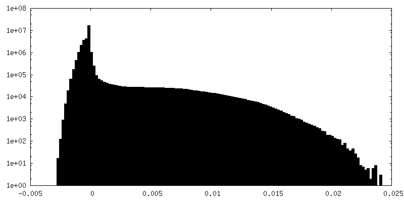
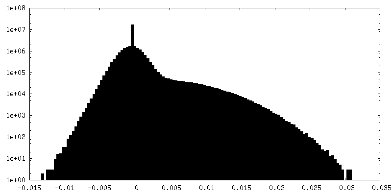
| Density |
| ||||||||||||||||||||||||||||||||||||
| Symmetry | Space group: 1 | ||||||||||||||||||||||||||||||||||||
| Details | EMDB XML:
|
 Z (Sec.)
 Y (Row.)
 X (Col.)


-Supplemental data
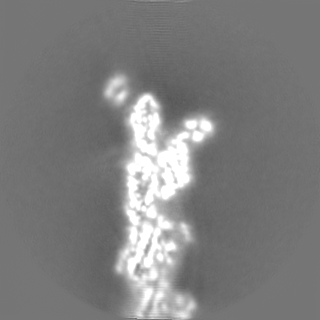
-Mask #1
| File | emd_19497_msk_1.map | ||||||||||||
|---|---|---|---|---|---|---|---|---|---|---|---|---|---|
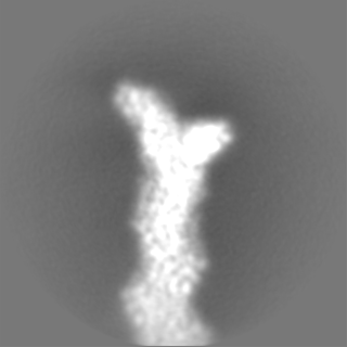
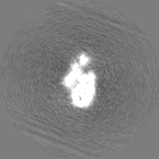
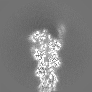
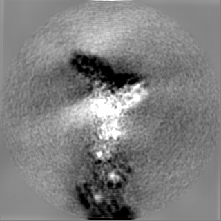
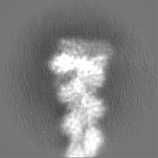
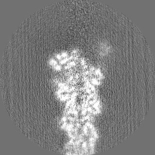
| Projections & Slices |
| ||||||||||||
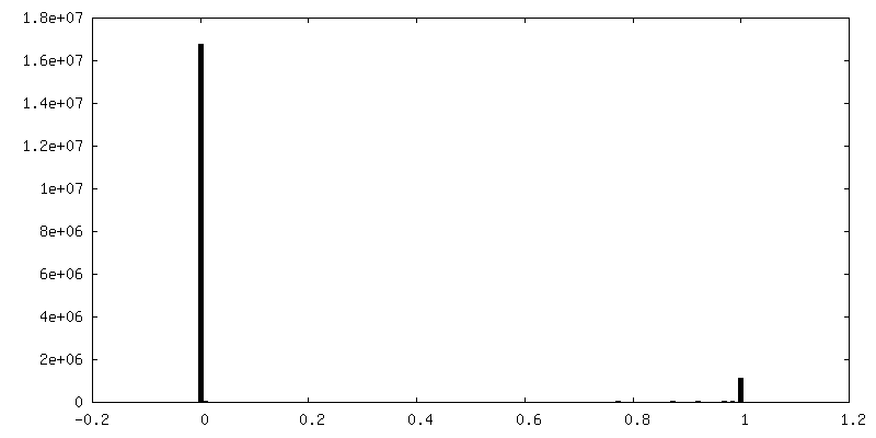
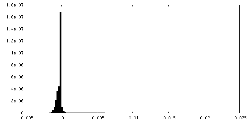
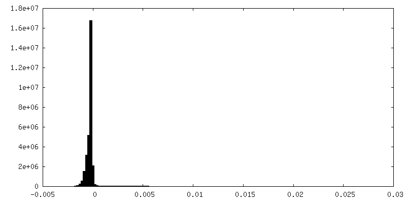
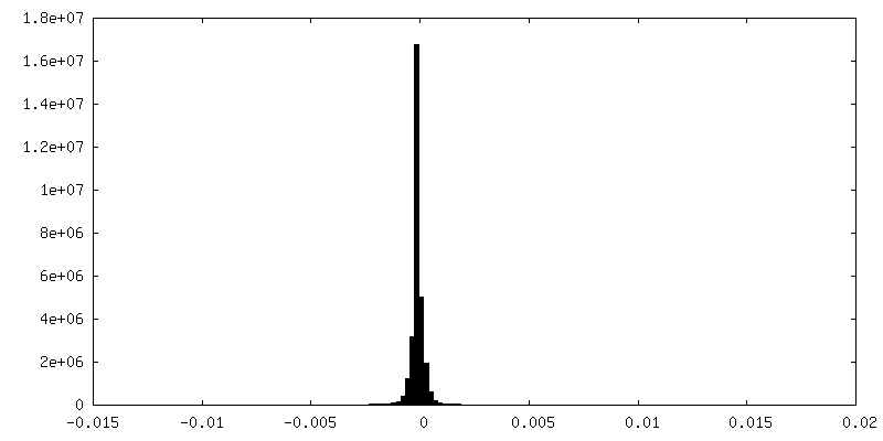
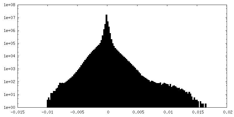
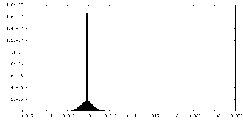
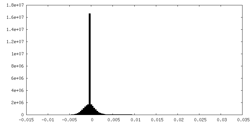
| Density Histograms |


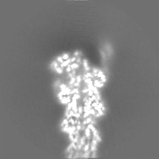
-Additional map: 3D-refined, unsharpened cryo-EM density map of the formin...
| File | emd_19497_additional_1.map | ||||||||||||
|---|---|---|---|---|---|---|---|---|---|---|---|---|---|
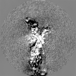
| Annotation | 3D-refined, unsharpened cryo-EM density map of the formin Cdc12 bound to the barbed end of F-actin (without phalloidin). | ||||||||||||
| Projections & Slices |
| ||||||||||||
| Density Histograms |


-Additional map: Cryo-EM map Cdc12 bound to phalloidin-stabilized F-actin (EMD-19496),...
| File | emd_19497_additional_2.map | ||||||||||||
|---|---|---|---|---|---|---|---|---|---|---|---|---|---|
| Annotation | Cryo-EM map Cdc12 bound to phalloidin-stabilized F-actin (EMD-19496), resampled on the cryo-EM reconstruction that was obtained without phalloidin. | ||||||||||||
| Projections & Slices |
| ||||||||||||
| Density Histograms |


-Additional map: Power-adjusted map of actin-Cdc12 with phalloidin(EMD-19496), resampled on...
| File | emd_19497_additional_3.map | ||||||||||||
|---|---|---|---|---|---|---|---|---|---|---|---|---|---|
| Annotation | Power-adjusted map of actin-Cdc12 with phalloidin(EMD-19496), resampled on the no-phalloidin map. Used to construct a difference map between the reconstructions with and without phalloidin. | ||||||||||||
| Projections & Slices |
| ||||||||||||
| Density Histograms |


-Additional map: Difference map between actin-Cdc12 reconstructions that were obtained...
| File | emd_19497_additional_4.map | ||||||||||||
|---|---|---|---|---|---|---|---|---|---|---|---|---|---|
| Annotation | Difference map between actin-Cdc12 reconstructions that were obtained in the absence (this entry) and presence (EMD-19496) of the toxin phalloidin. | ||||||||||||
| Projections & Slices |
| ||||||||||||
| Density Histograms |


-Half map: Unfiltered half map 1 of the formin Cdc12...
| File | emd_19497_half_map_1.map | ||||||||||||
|---|---|---|---|---|---|---|---|---|---|---|---|---|---|
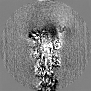
| Annotation | Unfiltered half map 1 of the formin Cdc12 bound to the barbed end of F-actin (without phalloidin). | ||||||||||||
| Projections & Slices |
| ||||||||||||
| Density Histograms |


-Half map: Unfiltered half map 2 of the formin Cdc12...
| File | emd_19497_half_map_2.map | ||||||||||||
|---|---|---|---|---|---|---|---|---|---|---|---|---|---|
| Annotation | Unfiltered half map 2 of the formin Cdc12 bound to the barbed end of F-actin (without phalloidin). | ||||||||||||
| Projections & Slices |
| ||||||||||||
| Density Histograms |


- Sample components
-Entire : Complex of the dimeric FH2 domain of S. Pombe Cdc12 bound to the ...
| Entire | Name: Complex of the dimeric FH2 domain of S. Pombe Cdc12 bound to the barbed end of F-actin. |
|---|---|
| Components |
|
-Supramolecule #1: Complex of the dimeric FH2 domain of S. Pombe Cdc12 bound to the ...
| Supramolecule | Name: Complex of the dimeric FH2 domain of S. Pombe Cdc12 bound to the barbed end of F-actin. type: complex / ID: 1 / Parent: 0 / Macromolecule list: all Details: Human beta-actin and S. Pombe Cdc12 were purified separately. Both proteins were mixed to assemble the complex prior to cryo-EM grid preparation. |
|---|---|
| Source (natural) | Organism: Homo sapiens (human) |
-Supramolecule #2: Actin filament
| Supramolecule | Name: Actin filament / type: complex / ID: 2 / Parent: 1 / Macromolecule list: #1 |
|---|
-Supramolecule #3: Dimeric FH2 domain of S. Pombe Cdc12
| Supramolecule | Name: Dimeric FH2 domain of S. Pombe Cdc12 / type: complex / ID: 3 / Parent: 1 / Macromolecule list: #2 |
|---|---|
| Source (natural) | Organism: Schizosaccharomyces pombe (fission yeast) |
-Macromolecule #1: human cytoplasmic beta-actin
| Macromolecule | Name: human cytoplasmic beta-actin / type: protein_or_peptide / ID: 1 Details: Actin purified recombinantly from BTI-Tnao38 cells. Enantiomer: LEVO |
|---|---|
| Source (natural) | Organism: Homo sapiens (human) |
| Recombinant expression | Organism:  Trichoplusia ni (cabbage looper) Trichoplusia ni (cabbage looper) |
| Sequence | String: MDDDIAALVV DNGSGMCKAG FAGDDAPRAV FPSIVGRPRH QGVMVGMGQK DSYVGDEAQS KRGILTLKYP IE(HIC)GIVTNWD DMEKIWHHTF YNELRVAPEE HPVLLTEAPL NPKANREKMT QIMFETFNTP AMYVAIQAVL SLYASGRTTG IVMDSGDGVT HTVPIYEGYA ...String: MDDDIAALVV DNGSGMCKAG FAGDDAPRAV FPSIVGRPRH QGVMVGMGQK DSYVGDEAQS KRGILTLKYP IE(HIC)GIVTNWD DMEKIWHHTF YNELRVAPEE HPVLLTEAPL NPKANREKMT QIMFETFNTP AMYVAIQAVL SLYASGRTTG IVMDSGDGVT HTVPIYEGYA LPHAILRLDL AGRDLTDYLM KILTERGYSF TTTAEREIVR DIKEKLCYVA LDFEQEMATA ASSSSLEKSY ELPDGQVITI GNERFRCPEA LFQPSFLGME SAGIHETTFN SIMKCDVDIR KDLYANTVLS GGTTMYPGIA DRMQKEITAL APSTMKIKII APPERKYSVW IGGSILASLS TFQQMWISKQ EYDESGPSIV HRKCF UniProtKB: Actin, cytoplasmic 1 |
-Macromolecule #2: S. pombe Cdc12
| Macromolecule | Name: S. pombe Cdc12 / type: protein_or_peptide / ID: 2 / Enantiomer: LEVO |
|---|---|
| Source (natural) | Organism: Schizosaccharomyces pombe (fission yeast) |
| Recombinant expression | Organism:  Escherichia coli (E. coli) Escherichia coli (E. coli) |
| Sequence | String: SKDDLHKTTG LTRRPTRRLK QMHWEKLNSG LEFTFWTGPS DEANKILETL HTSGVLDELD ESFAMKEAK TLVKKTCART DYMSSELQKL FGIHFHKLSH KNPNEIIRMI LHCDDSMNEC V EFLSSDKV LNQPKLKADL EPYRIDWANG GDLVNSEKDA SELSRWDYLY ...String: SKDDLHKTTG LTRRPTRRLK QMHWEKLNSG LEFTFWTGPS DEANKILETL HTSGVLDELD ESFAMKEAK TLVKKTCART DYMSSELQKL FGIHFHKLSH KNPNEIIRMI LHCDDSMNEC V EFLSSDKV LNQPKLKADL EPYRIDWANG GDLVNSEKDA SELSRWDYLY VRLIVDLGGY WN QRMNALK VKNIIETNYE NLVRQTKLIG RAALELRDSK VFKGLLYLIL YLGNYMNDYV RQA KGFAIG SLQRLPLIKN ANNTKSLLHI LDITIRKHFP QFDNFSPELS TVTEAAKLNI EAIE QECSE LIRGCQNLQI DCDSGALSDP TVFHPDDKIL SVILPWLMEG TKKMDFLKEH LRTMN TTLN NAMRYFGEQP NDPNSKNLFF KRVDSFIIDY SKARSDNLKS EEEEASQHRR LNLVN UniProtKB: Cell division control protein 12 |
-Experimental details
-Structure determination
| Method | cryo EM |
|---|---|
 Processing Processing | single particle reconstruction |
| Aggregation state | particle |
-Sample preparation
| Buffer | pH: 7.1 Component:
Details: 1xKMEH (10 mM HEPES pH 7.1, 100 mM KCl, 2 mM MgCl2, 1 mM EGTA, 0.5 mM TCEP) | ||||||||||||||||||
|---|---|---|---|---|---|---|---|---|---|---|---|---|---|---|---|---|---|---|---|
| Grid | Model: Quantifoil R2/1 / Material: GOLD / Mesh: 200 / Support film - Material: CARBON / Support film - topology: HOLEY / Pretreatment - Type: GLOW DISCHARGE / Pretreatment - Time: 90 sec. | ||||||||||||||||||
| Vitrification | Cryogen name: ETHANE-PROPANE / Chamber humidity: 100 % / Chamber temperature: 286 K / Instrument: FEI VITROBOT MARK IV / Details: 3 seconds, force 0.. |
- Electron microscopy
| Microscope | FEI TITAN KRIOS |
|---|---|
| Electron beam | Acceleration voltage: 300 kV / Electron source: FIELD EMISSION GUN |
| Electron optics | C2 aperture diameter: 50.0 µm / Illumination mode: FLOOD BEAM / Imaging mode: BRIGHT FIELDBright-field microscopy / Cs: 0.01 mm / Nominal defocus max: 2.7 µm / Nominal defocus min: 1.2 µm / Nominal magnification: 81000 |
| Specialist optics | Spherical aberration corrector: Titan Krios G2 microscope (Thermo Fisher Scientific) with an in-column Cs-corrector. Energy filter - Name: GIF Bioquantum / Energy filter - Slit width: 15 eV / Details: Gatan energy filter. |
| Sample stage | Specimen holder model: FEI TITAN KRIOS AUTOGRID HOLDER / Cooling holder cryogen: NITROGEN |
| Details | 300 kV Titan Krios G2 microscope (Thermo Fisher Scientific) with an in-column Cs-corrector. |
| Image recording | Film or detector model: GATAN K3 BIOQUANTUM (6k x 4k) / Number grids imaged: 1 / Number real images: 12009 / Average electron dose: 64.3 e/Å2 |
| Experimental equipment |  Model: Titan Krios / Image courtesy: FEI Company |
-Image processing
| Particle selection | Number selected: 2443647 / Details: Particles picked using SPHIRE-crYOLO. |
|---|---|
| Startup model | Type of model: EMDB MAP EMDB ID: Details: Cdc12 bound to phalloidin-stabilized F-actin. |
| Initial angle assignment | Type: MAXIMUM LIKELIHOOD / Software - Name: cryoSPARC (ver. v3.3.2) |
| Final 3D classification | Number classes: 4 / Software - Name: RELION (ver. 3.1.0) / Details: 3D classification in RELION. |
| Final angle assignment | Type: MAXIMUM LIKELIHOOD / Software - Name: RELION (ver. 3.1.0) |
| Final reconstruction | Applied symmetry - Point group: C1 (asymmetric) / Resolution.type: BY AUTHOR / Resolution: 4.54 Å / Resolution method: FSC 0.143 CUT-OFF / Software - Name: RELION (ver. 3.1.0) Details: Final reconstruction displays preferred orientation. Number images used: 173582 |
| FSC plot (resolution estimation) | 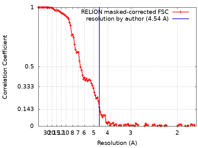 |
-Atomic model buiding 1
| Initial model | PDB ID: Chain - Source name: PDB / Chain - Initial model type: experimental model |
|---|