 Movie
Movie Controller

[English] 日本語
 Yorodumi
- PDB-8ced: Rnase R bound to a 30S degradation intermediate (State I - head-t... -
+ Open data

- Basic information
| Entry | Database: PDB / ID: 8ced | ||||||
|---|---|---|---|---|---|---|---|
| Title | Rnase R bound to a 30S degradation intermediate (State I - head-turning) | ||||||
 Components Components |
| ||||||
 Keywords Keywords |  RIBOSOME / SSU / 30S / RNase R / ribosomal degradation / turnover RIBOSOME / SSU / 30S / RNase R / ribosomal degradation / turnover | ||||||
| Function / homology |  Function and homology informationribosomal small subunit biogenesis / small ribosomal subunit rRNA binding / ribosomal small subunit assembly / cytosolic small ribosomal subunit / small ribosomal subunit / tRNA binding / rRNA binding / ribosome / structural constituent of ribosome / ribonucleoprotein complex ...ribosomal small subunit biogenesis / small ribosomal subunit rRNA binding / ribosomal small subunit assembly / cytosolic small ribosomal subunit / small ribosomal subunit / tRNA binding / rRNA binding / ribosome / structural constituent of ribosome / ribonucleoprotein complex / translation / response to antibiotic / mRNA binding / RNA binding / zinc ion binding / cytosol / cytoplasm Function and homology informationribosomal small subunit biogenesis / small ribosomal subunit rRNA binding / ribosomal small subunit assembly / cytosolic small ribosomal subunit / small ribosomal subunit / tRNA binding / rRNA binding / ribosome / structural constituent of ribosome / ribonucleoprotein complex ...ribosomal small subunit biogenesis / small ribosomal subunit rRNA binding / ribosomal small subunit assembly / cytosolic small ribosomal subunit / small ribosomal subunit / tRNA binding / rRNA binding / ribosome / structural constituent of ribosome / ribonucleoprotein complex / translation / response to antibiotic / mRNA binding / RNA binding / zinc ion binding / cytosol / cytoplasmSimilarity search - Function | ||||||
| Biological species |  Bacillus subtilis subsp. subtilis str. 168 (bacteria) Bacillus subtilis subsp. subtilis str. 168 (bacteria) | ||||||
| Method | ELECTRON MICROSCOPY / single particle reconstruction / cryo EM / Resolution: 4.15 Å | ||||||
 Authors Authors | Paternoga, H. / Dimitrova-Paternoga, L. / Wilson, D.N. | ||||||
| Funding support |  Germany, 1items Germany, 1items
| ||||||
 Citation Citation | Journal: Nature / Year: 2024 Title: Structural basis of ribosomal 30S subunit degradation by RNase R. Authors: Lyudmila Dimitrova-Paternoga / Sergo Kasvandik / Bertrand Beckert / Sander Granneman / Tanel Tenson / Daniel N Wilson / Helge Paternoga /    Abstract: Protein synthesis is a major energy-consuming process of the cell that requires the controlled production and turnover of ribosomes. Although the past few years have seen major advances in our ...Protein synthesis is a major energy-consuming process of the cell that requires the controlled production and turnover of ribosomes. Although the past few years have seen major advances in our understanding of ribosome biogenesis, structural insight into the degradation of ribosomes has been lacking. Here we present native structures of two distinct small ribosomal 30S subunit degradation intermediates associated with the 3' to 5' exonuclease ribonuclease R (RNase R). The structures reveal that RNase R binds at first to the 30S platform to facilitate the degradation of the functionally important anti-Shine-Dalgarno sequence and the decoding-site helix 44. RNase R then encounters a roadblock when it reaches the neck region of the 30S subunit, and this is overcome by a major structural rearrangement of the 30S head, involving the loss of ribosomal proteins. RNase R parallels this movement and relocates to the decoding site by using its N-terminal helix-turn-helix domain as an anchor. In vitro degradation assays suggest that head rearrangement poses a major kinetic barrier for RNase R, but also indicate that the enzyme alone is sufficient for complete degradation of 30S subunits. Collectively, our results provide a mechanistic basis for the degradation of 30S mediated by RNase R, and reveal that RNase R targets orphaned 30S subunits using a dynamic mechanism involving an anchored switching of binding sites. | ||||||
| History |
|
- Structure visualization
| Structure viewer | Molecule: MolmilJmol/JSmol |
|---|
- Downloads & links
-Download
| PDBx/mmCIF format | 8ced.cif.gz | 1.3 MB | Display | PDBx/mmCIF format |
|---|---|---|---|---|
| PDB format | pdb8ced.ent.gz | 1 MB | Display | PDB format |
| PDBx/mmJSON format | 8ced.json.gz | Tree view | PDBx/mmJSON format | |
| Others |  Other downloads Other downloads |
-Validation report
| Arichive directory | https://data.pdbj.org/pub/pdb/validation_reports/ce/8cedftp://data.pdbj.org/pub/pdb/validation_reports/ce/8ced | HTTPS FTP |
|---|
-Related structure data
| Related structure data |  16606MC 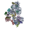 8cduC 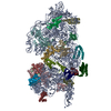 8cdvC 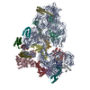 8cecC 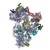 8ceeC C: citing same article ( M: map data used to model this data |
|---|---|
| Similar structure data |


-Links
 PDBj
PDBj


- Assembly
| Deposited unit | 
|
|---|---|
| 1 |
|
-Components
-RNA chain , 2 types, 2 molecules AB
| #1: RNA chain | Mass: 503369.125 Da / Num. of mol.: 1 / Source method: isolated from a natural source Source: (natural) Bacillus subtilis subsp. subtilis str. 168 (bacteria)References: GenBank: 225184640 |
|---|---|
| #2: RNA chain | Mass: 2259.483 Da / Num. of mol.: 1 / Source method: isolated from a natural source Source: (natural) Bacillus subtilis subsp. subtilis str. 168 (bacteria) |
-Protein , 1 types, 1 molecules C
| #3: Protein | Mass: 88886.312 Da / Num. of mol.: 1 / Source method: isolated from a natural source Source: (natural) Bacillus subtilis subsp. subtilis str. 168 (bacteria) |
|---|
-30S ribosomal protein ... , 19 types, 19 molecules DFGILOPQSTUVEHJKMNR
| #4: Protein | / BS1 / Vegetative protein 209 / VEG209 Mass: 28009.297 Da / Num. of mol.: 1 / Source method: isolated from a natural source Source: (natural) Bacillus subtilis subsp. subtilis str. 168 (bacteria)References: UniProt: P21464 |
|---|---|
| #5: Protein | / BS4 Mass: 22874.271 Da / Num. of mol.: 1 / Source method: isolated from a natural source Source: (natural) Bacillus subtilis subsp. subtilis str. 168 (bacteria)References: UniProt: P21466 |
| #6: Protein | / BS5 Mass: 17650.625 Da / Num. of mol.: 1 / Source method: isolated from a natural source Source: (natural) Bacillus subtilis subsp. subtilis str. 168 (bacteria)References: UniProt: P21467 |
| #7: Protein | / BS8 Mass: 14901.427 Da / Num. of mol.: 1 / Source method: isolated from a natural source Source: (natural) Bacillus subtilis subsp. subtilis str. 168 (bacteria)References: UniProt: P12879 |
| #8: Protein | / BS12 Mass: 15248.736 Da / Num. of mol.: 1 / Source method: isolated from a natural source Source: (natural) Bacillus subtilis subsp. subtilis str. 168 (bacteria)References: UniProt: P21472 |
| #9: Protein | / BS18 Mass: 10597.224 Da / Num. of mol.: 1 / Source method: isolated from a natural source Source: (natural) Bacillus subtilis subsp. subtilis str. 168 (bacteria)References: UniProt: P21473 |
| #10: Protein | / BS17 Mass: 10153.833 Da / Num. of mol.: 1 / Source method: isolated from a natural source Source: (natural) Bacillus subtilis subsp. subtilis str. 168 (bacteria)References: UniProt: P21474 |
| #11: Protein | / BS16 Mass: 10220.979 Da / Num. of mol.: 1 / Source method: isolated from a natural source Source: (natural) Bacillus subtilis subsp. subtilis str. 168 (bacteria)References: UniProt: P12874 |
| #12: Protein | / BS20 Mass: 9622.217 Da / Num. of mol.: 1 / Source method: isolated from a natural source Source: (natural) Bacillus subtilis subsp. subtilis str. 168 (bacteria)References: UniProt: P21477 |
| #13: Protein | / BS9 Mass: 11140.548 Da / Num. of mol.: 1 / Source method: isolated from a natural source Source: (natural) Bacillus subtilis subsp. subtilis str. 168 (bacteria)References: UniProt: P21468 |
| #14: Protein | / BS21 Mass: 8990.613 Da / Num. of mol.: 1 / Source method: isolated from a natural source Source: (natural) Bacillus subtilis subsp. subtilis str. 168 (bacteria)References: UniProt: P21475 |
| #15: Protein | / BS11 Mass: 13952.000 Da / Num. of mol.: 1 / Source method: isolated from a natural source Source: (natural) Bacillus subtilis subsp. subtilis str. 168 (bacteria)References: UniProt: P04969 |
| #16: Protein | / BS3 / BS2 Mass: 24364.887 Da / Num. of mol.: 1 / Source method: isolated from a natural source Source: (natural) Bacillus subtilis subsp. subtilis str. 168 (bacteria)References: UniProt: P21465 |
| #17: Protein | / BS7 Mass: 17915.879 Da / Num. of mol.: 1 / Source method: isolated from a natural source Source: (natural) Bacillus subtilis subsp. subtilis str. 168 (bacteria)References: UniProt: P21469 |
| #18: Protein | / BS10 Mass: 14335.504 Da / Num. of mol.: 1 / Source method: isolated from a natural source Source: (natural) Bacillus subtilis subsp. subtilis str. 168 (bacteria)References: UniProt: P21470 |
| #19: Protein | / BS13 Mass: 11687.661 Da / Num. of mol.: 1 / Source method: isolated from a natural source Source: (natural) Bacillus subtilis subsp. subtilis str. 168 (bacteria)References: UniProt: P21471 |
| #20: Protein | / BS14 Mass: 13818.085 Da / Num. of mol.: 1 / Source method: isolated from a natural source Source: (natural) Bacillus subtilis subsp. subtilis str. 168 (bacteria)References: UniProt: P20282 |
| #21: Protein | / 30S ribosomal protein S14 type Z / 30S ribosomal protein S14-1 / BS-A Mass: 7263.803 Da / Num. of mol.: 1 / Source method: isolated from a natural source Source: (natural) Bacillus subtilis subsp. subtilis str. 168 (bacteria)References: UniProt: P12878 |
| #22: Protein | / BS19 Mass: 10607.309 Da / Num. of mol.: 1 / Source method: isolated from a natural source Source: (natural) Bacillus subtilis subsp. subtilis str. 168 (bacteria)References: UniProt: P21476 |
-Experimental details
-Experiment
| Experiment | Method: ELECTRON MICROSCOPY |
|---|---|
| EM experiment | Aggregation state: PARTICLE / 3D reconstruction method: single particle reconstruction |
- Sample preparation
| Component | Name: 30S ribosomal subunit in complex with 3' exonuclease RNase R Type: RIBOSOME / Entity ID: all / Source: NATURAL |
|---|---|
| Molecular weight | Experimental value: NO |
| Source (natural) | Organism: Bacillus subtilis subsp. subtilis str. 168 (bacteria) |
| Buffer solution | pH: 7.5 |
| Specimen | Embedding applied: NO / Shadowing applied: NO / Staining applied: NO / Vitrification applied: YES |
| Vitrification | Cryogen name: ETHANE-PROPANE |
- Electron microscopy imaging
| Experimental equipment |  Model: Titan Krios / Image courtesy: FEI Company |
|---|---|
| Microscopy | Model: FEI TITAN KRIOS |
| Electron gun | Electron source: FIELD EMISSION GUN / Accelerating voltage: 300 kV / Illumination mode: FLOOD BEAM |
| Electron lens | Mode: BRIGHT FIELDBright-field microscopy / Nominal defocus max: 900 nm / Nominal defocus min: 400 nm |
| Image recording | Electron dose: 50 e/Å2 / Film or detector model: FEI FALCON IV (4k x 4k) |
- Processing
| Software | Name: REFMAC / Version: 5.8.0403 / Classification: refinement | ||||||||||||||||||||||||||||||||||||||||||||||||||||||||||||||||||||||||||||||||||||||||||||||||||||||||||
|---|---|---|---|---|---|---|---|---|---|---|---|---|---|---|---|---|---|---|---|---|---|---|---|---|---|---|---|---|---|---|---|---|---|---|---|---|---|---|---|---|---|---|---|---|---|---|---|---|---|---|---|---|---|---|---|---|---|---|---|---|---|---|---|---|---|---|---|---|---|---|---|---|---|---|---|---|---|---|---|---|---|---|---|---|---|---|---|---|---|---|---|---|---|---|---|---|---|---|---|---|---|---|---|---|---|---|---|
| EM software |
| ||||||||||||||||||||||||||||||||||||||||||||||||||||||||||||||||||||||||||||||||||||||||||||||||||||||||||
| CTF correction | Type: PHASE FLIPPING AND AMPLITUDE CORRECTION | ||||||||||||||||||||||||||||||||||||||||||||||||||||||||||||||||||||||||||||||||||||||||||||||||||||||||||
| Particle selection | Num. of particles selected: 2303673 | ||||||||||||||||||||||||||||||||||||||||||||||||||||||||||||||||||||||||||||||||||||||||||||||||||||||||||
| 3D reconstruction | Resolution: 4.15 Å / Resolution method: FSC 0.143 CUT-OFF / Num. of particles: 6540 / Symmetry type: POINT | ||||||||||||||||||||||||||||||||||||||||||||||||||||||||||||||||||||||||||||||||||||||||||||||||||||||||||
| Refinement | Resolution: 4.15→232 Å / Cor.coef. Fo:Fc: 0.791 / SU B: 59.691 / SU ML: 0.742 / ESU R: 1.321 Stereochemistry target values: MAXIMUM LIKELIHOOD WITH PHASES Details: HYDROGENS HAVE BEEN ADDED IN THE RIDING POSITIONS
| ||||||||||||||||||||||||||||||||||||||||||||||||||||||||||||||||||||||||||||||||||||||||||||||||||||||||||
| Solvent computation | Solvent model: PARAMETERS FOR MASK CACLULATION | ||||||||||||||||||||||||||||||||||||||||||||||||||||||||||||||||||||||||||||||||||||||||||||||||||||||||||
| Displacement parameters | Biso mean: 230.775 Å2 | ||||||||||||||||||||||||||||||||||||||||||||||||||||||||||||||||||||||||||||||||||||||||||||||||||||||||||
| Refinement step | Cycle: 1 / Total: 51964 | ||||||||||||||||||||||||||||||||||||||||||||||||||||||||||||||||||||||||||||||||||||||||||||||||||||||||||
| Refine LS restraints |
|