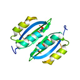
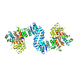
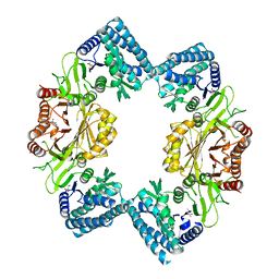
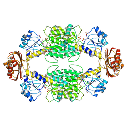
Loading
PDBj
MenuPDBj@FacebookPDBj@TwitterPDBj@YouTubewwPDB FoundationwwPDB
RCSB PDBPDBeBMRBAdv. SearchSearch help

2LVW
DownloadVisualize
BU of 2lvw by Molmil
Solution NMR studies of the dimeric regulatory subunit IlvN of the E.coli Acetohydroxyacid synthase I (AHAS I)
Descriptor: Acetolactate synthase isozyme 1 small subunit
Authors:Karanth, M.N, Sarma, S.P.
Deposit date:2012-07-12
Release date:2013-05-29
Last modified:2024-05-01
Method:SOLUTION NMR
Cite:The coil-to-helix transition in IlvN regulates the allosteric control of Escherichia coli acetohydroxyacid synthase I
Biochemistry, 52, 2013
3TVI
DownloadVisualize
BU of 3tvi by Molmil
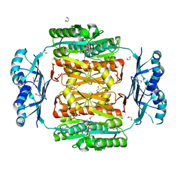
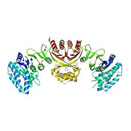
Crystal structure of Clostridium acetobutylicum aspartate kinase (CaAK): An important allosteric enzyme for industrial amino acids production
Descriptor: ASPARTIC ACID, Aspartokinase, LYSINE
Authors:Manjasetty, B.A, Chance, M.R, Burley, S.K, Almo, S.C, New York SGX Research Center for Structural Genomics (NYSGXRC)
Deposit date:2011-09-20
Release date:2011-11-09
Last modified:2021-02-10
Method:X-RAY DIFFRACTION (3 Å)
Cite:Crystal structure of Clostridium acetobutylicum Aspartate kinase (CaAK): An important allosteric enzyme for amino acids production.
Biotechnol Rep (Amst), 3, 2014
3S1T
DownloadVisualize
BU of 3s1t by Molmil
Structure of the regulatory domain of aspartokinase (Rv3709c; AK-beta) in complex with threonine from Mycobacterium tuberculosis
Descriptor: Aspartokinase, SULFATE ION, THREONINE
Authors:Schuldt, L, Mueller-Dieckmann, J, Weiss, M.S.
Deposit date:2011-05-16
Release date:2012-06-13
Last modified:2023-09-13
Method:X-RAY DIFFRACTION (1.63 Å)
Cite:High-resolution X-ray structure of the regulatory domain of the mycobacterial aspartokinase in complex with threonine
To be Published
3OBI
DownloadVisualize
BU of 3obi by Molmil
Crystal structure of a formyltetrahydrofolate deformylase (NP_949368) from RHODOPSEUDOMONAS PALUSTRIS CGA009 at 1.95 A resolution
Descriptor: 1,2-ETHANEDIOL, Formyltetrahydrofolate deformylase
Authors:Joint Center for Structural Genomics (JCSG)
Deposit date:2010-08-06
Release date:2010-08-18
Last modified:2023-02-01
Method:X-RAY DIFFRACTION (1.95 Å)
Cite:Crystal structure of a formyltetrahydrofolate deformylase (NP_949368) from RHODOPSEUDOMONAS PALUSTRIS CGA009 at 1.95 A resolution
To be published
3NRB
DownloadVisualize
BU of 3nrb by Molmil
Crystal structure of a formyltetrahydrofolate deformylase (purU, PP_1943) from PSEUDOMONAS PUTIDA KT2440 at 2.05 A resolution
Descriptor: 1,2-ETHANEDIOL, CITRATE ANION, Formyltetrahydrofolate deformylase, ...
Authors:Joint Center for Structural Genomics (JCSG)
Deposit date:2010-06-30
Release date:2010-07-28
Last modified:2023-02-01
Method:X-RAY DIFFRACTION (2.05 Å)
Cite:Crystal structure of a formyltetrahydrofolate deformylase (purU, PP_1943) from PSEUDOMONAS PUTIDA KT2440 at 2.05 A resolution
To be published
3MWB
DownloadVisualize
BU of 3mwb by Molmil
The Crystal Structure of Prephenate dehydratase in complex with L-Phe from Arthrobacter aurescens to 2.0A
Descriptor: MAGNESIUM ION, PHENYLALANINE, Prephenate dehydratase
Authors:Stein, A.J, Chhor, G, Buck, K, Joachimiak, A, Midwest Center for Structural Genomics (MCSG)
Deposit date:2010-05-05
Release date:2010-06-30
Last modified:2017-11-08
Method:X-RAY DIFFRACTION (2 Å)
Cite:The Crystal Structure of Prephenate dehydratase in complex with L-Phe from Arthrobacter aurescens to 2.0A
To be Published
3MTJ
DownloadVisualize
BU of 3mtj by Molmil
The Crystal Structure of a Homoserine Dehydrogenase from Thiobacillus denitrificans to 2.15A
Descriptor: Homoserine dehydrogenase, SULFATE ION
Authors:Stein, A.J, Cui, H, Chin, S, Savchenko, A, Joachimiak, A, Midwest Center for Structural Genomics (MCSG)
Deposit date:2010-04-30
Release date:2010-05-12
Last modified:2017-11-08
Method:X-RAY DIFFRACTION (2.15 Å)
Cite:The Crystal Structure of a Homoserine Dehydrogenase from Thiobacillus denitrificans to 2.15A
To be Published
3L76
DownloadVisualize
BU of 3l76 by Molmil
Crystal Structure of Aspartate Kinase from Synechocystis
Descriptor: Aspartokinase, LYSINE, SULFATE ION, ...
Authors:Robin, A, Cobessi, D, Curien, G, Robert-Genthon, M, Ferrer, J.-L, Dumas, R.
Deposit date:2009-12-28
Release date:2010-06-09
Last modified:2024-03-20
Method:X-RAY DIFFRACTION (2.54 Å)
Cite:A new mode of dimerization of allosteric enzymes with ACT domains revealed by the crystal structure of the aspartate kinase from Cyanobacteria
J.Mol.Biol., 399, 2010
3AB2
DownloadVisualize
BU of 3ab2 by Molmil
Crystal structure of aspartate kinase from Corynebacterium glutamicum in complex with threonine
Descriptor: Aspartokinase, THREONINE
Authors:Yoshida, A, Tomita, T, Kuzuyama, T, Nishiyama, M.
Deposit date:2009-11-30
Release date:2010-06-23
Last modified:2023-11-01
Method:X-RAY DIFFRACTION (2.59 Å)
Cite:Mechanism of concerted inhibition of {alpha}2{beta}2-type heterooligomeric aspartate kinase from Corynebacterium glutamicum
J.Biol.Chem., 285, 2010
3AB4
DownloadVisualize
BU of 3ab4 by Molmil
Crystal structure of feedback inhibition resistant mutant of aspartate kinase from Corynebacterium glutamicum in complex with lysine and threonine
Descriptor: Aspartokinase, LYSINE, THREONINE
Authors:Yoshida, A, Tomita, T, Kuzuyama, T, Nishiyama, M.
Deposit date:2009-11-30
Release date:2010-06-23
Last modified:2023-11-01
Method:X-RAY DIFFRACTION (2.47 Å)
Cite:Mechanism of concerted inhibition of {alpha}2{beta}2-type heterooligomeric aspartate kinase from Corynebacterium glutamicum
J.Biol.Chem., 285, 2010
3AAW
DownloadVisualize
BU of 3aaw by Molmil
Crystal structure of aspartate kinase from Corynebacterium glutamicum in complex with lysine and threonine
Descriptor: Aspartokinase, Aspartokinase LysC beta subunit, LYSINE, ...
Authors:Yoshida, A, Tomita, T, Kuzuyama, T, Nishiyama, M.
Deposit date:2009-11-27
Release date:2010-06-23
Last modified:2023-11-01
Method:X-RAY DIFFRACTION (2.5 Å)
Cite:Mechanism of concerted inhibition of {alpha}2{beta}2-type heterooligomeric aspartate kinase from Corynebacterium glutamicum
J.Biol.Chem., 285, 2010
3DDN
DownloadVisualize
BU of 3ddn by Molmil
Crystal structure of hydroxypyruvic acid phosphate bound D-3-phosphoglycerate dehydrogenase in mycobacterium tuberculosis
Descriptor: 2-oxo-3-(phosphonooxy)propanoic acid, D-3-phosphoglycerate dehydrogenase, L(+)-TARTARIC ACID
Authors:Dey, S, Sacchettini, J.C.
Deposit date:2008-06-05
Release date:2008-07-22
Last modified:2023-11-01
Method:X-RAY DIFFRACTION (2.4 Å)
Cite:Structural analysis of substrate and effector binding in Mycobacterium tuberculosis D-3-phosphoglycerate dehydrogenase
Biochemistry, 47, 2008
3DC2
DownloadVisualize
BU of 3dc2 by Molmil
Crystal structure of serine bound D-3-phosphoglycerate dehydrogenase from Mycobacterium tuberculosis
Descriptor: D-3-phosphoglycerate dehydrogenase, L(+)-TARTARIC ACID, SERINE
Authors:Dey, S, Sacchettini, J.C.
Deposit date:2008-06-03
Release date:2008-07-22
Last modified:2023-11-01
Method:X-RAY DIFFRACTION (2.7 Å)
Cite:Structural analysis of substrate and effector binding in Mycobacterium tuberculosis D-3-phosphoglycerate dehydrogenase
Biochemistry, 47, 2008
2ZHO
DownloadVisualize
BU of 2zho by Molmil
Crystal structure of the regulatory subunit of aspartate kinase from Thermus thermophilus (ligand free form)
Descriptor: Aspartokinase
Authors:Yoshida, A, Tomita, T, Kuzuyama, T, Nishiyama, M.
Deposit date:2008-02-06
Release date:2009-02-17
Last modified:2023-11-01
Method:X-RAY DIFFRACTION (2.98 Å)
Cite:Crystal structures of the regulatory subunit of Thr-sensitive aspartate kinase from Thermus thermophilus
Febs J., 276, 2009
3C20
DownloadVisualize
BU of 3c20 by Molmil
Crystal Structure of Threonine-sensitive Aspartokinase from Methanococcus jannaschii with L-aspartate
Descriptor: ASPARTIC ACID, FORMIC ACID, Probable aspartokinase
Authors:Liu, X, Pavlovsky, A.G, Viola, R.E.
Deposit date:2008-01-24
Release date:2008-04-29
Last modified:2023-08-30
Method:X-RAY DIFFRACTION (2.7 Å)
Cite:The Structural Basis for Allosteric Inhibition of a Threonine-sensitive Aspartokinase.
J.Biol.Chem., 283, 2008
3C1N
DownloadVisualize
BU of 3c1n by Molmil
Crystal Structure of Allosteric Inhibition Threonine-sensitive Aspartokinase from Methanococcus jannaschii with L-threonine
Descriptor: Probable aspartokinase, THREONINE
Authors:Liu, X, Pavlovshy, A.G, Viola, R.E.
Deposit date:2008-01-23
Release date:2008-03-25
Last modified:2023-08-30
Method:X-RAY DIFFRACTION (2.72 Å)
Cite:The Structural Basis for Allosteric Inhibition of a Threonine-sensitive Aspartokinase.
J.Biol.Chem., 283, 2008
3C1M
DownloadVisualize
BU of 3c1m by Molmil
Cyrstal Structure of threonine-sensitive aspartokinase from Methanococcus jannaschii with MgAMP-PNP and L-aspartate
Descriptor: ASPARTIC ACID, FORMIC ACID, MAGNESIUM ION, ...
Authors:Liu, X.
Deposit date:2008-01-23
Release date:2008-03-25
Last modified:2024-02-21
Method:X-RAY DIFFRACTION (2.3 Å)
Cite:The Structural Basis for Allosteric Inhibition of a Threonine-sensitive Aspartokinase.
J.Biol.Chem., 283, 2008
2RE1
DownloadVisualize
BU of 2re1 by Molmil
Crystal structure of aspartokinase alpha and beta subunits
Descriptor: Aspartokinase, alpha and beta subunits, CALCIUM ION
Authors:Chang, C, Li, H, Gu, M, Joachimiak, A, Midwest Center for Structural Genomics (MCSG)
Deposit date:2007-09-25
Release date:2007-10-09
Last modified:2011-07-13
Method:X-RAY DIFFRACTION (2.75 Å)
Cite:Crystal structure of aspartokinase alpha and beta subunits.
To be Published
2QMW
DownloadVisualize
BU of 2qmw by Molmil
The crystal structure of the prephenate dehydratase (PDT) from Staphylococcus aureus subsp. aureus Mu50
Descriptor: 1,2-ETHANEDIOL, ACETATE ION, DI(HYDROXYETHYL)ETHER, ...
Authors:Tan, K, Zhang, R, Li, H, Gu, M, Joachimiak, A, Midwest Center for Structural Genomics (MCSG)
Deposit date:2007-07-17
Release date:2007-08-07
Last modified:2011-07-13
Method:X-RAY DIFFRACTION (2.3 Å)
Cite:Structures of open (R) and close (T) states of prephenate dehydratase (PDT) - implication of allosteric regulation by L-phenylalanine.
J.Struct.Biol., 162, 2008
2QMX
DownloadVisualize
BU of 2qmx by Molmil
The crystal structure of L-Phe inhibited prephenate dehydratase from Chlorobium tepidum TLS
Descriptor: 1,2-ETHANEDIOL, ACETATE ION, PHENYLALANINE, ...
Authors:Tan, K, Li, H, Clancy, S, Joachimiak, A, Midwest Center for Structural Genomics (MCSG)
Deposit date:2007-07-17
Release date:2007-08-07
Last modified:2011-07-13
Method:X-RAY DIFFRACTION (2.3 Å)
Cite:Structures of open (R) and close (T) states of prephenate dehydratase (PDT) - implication of allosteric regulation by L-phenylalanine.
J.Struct.Biol., 162, 2008
2PA3
DownloadVisualize
BU of 2pa3 by Molmil
crystal structure of serine bound G336V mutant of E.coli phosphoglycerate dehydrogenase
Descriptor: 1,4-DIHYDRONICOTINAMIDE ADENINE DINUCLEOTIDE, D-3-phosphoglycerate dehydrogenase, SERINE
Authors:Dey, S, Sacchettini, J.C.
Deposit date:2007-03-27
Release date:2007-04-24
Last modified:2023-08-30
Method:X-RAY DIFFRACTION (2.74 Å)
Cite:The Effect of Hinge Mutations on Effector Binding and Domain Rotation in Escherichia coli D-3-Phosphoglycerate Dehydrogenase.
J.Biol.Chem., 282, 2007
2P9G
DownloadVisualize
BU of 2p9g by Molmil
Crystal structure of serine bound G336V,G337V double mutant of E.coli phosphoglycerate dehydrogenase
Descriptor: 1,4-DIHYDRONICOTINAMIDE ADENINE DINUCLEOTIDE, D-3-phosphoglycerate dehydrogenase, SERINE
Authors:Dey, S, Sacchettini, J.C.
Deposit date:2007-03-25
Release date:2007-04-24
Last modified:2023-08-30
Method:X-RAY DIFFRACTION (2.8 Å)
Cite:The Effect of Hinge Mutations on Effector Binding and Domain Rotation in Escherichia coli D-3-Phosphoglycerate Dehydrogenase.
J.Biol.Chem., 282, 2007
2P9E
DownloadVisualize
BU of 2p9e by Molmil
Crystal Structure of G336V mutant of E.coli phosphoglycerate dehydrogenase
Descriptor: 1,4-DIHYDRONICOTINAMIDE ADENINE DINUCLEOTIDE, CITRIC ACID, D-3-phosphoglycerate dehydrogenase, ...
Authors:Dey, S, Sacchettini, J.C.
Deposit date:2007-03-25
Release date:2007-04-24
Last modified:2023-08-30
Method:X-RAY DIFFRACTION (2.6 Å)
Cite:The Effect of Hinge Mutations on Effector Binding and Domain Rotation in Escherichia coli D-3-Phosphoglycerate Dehydrogenase
J.Biol.Chem., 282, 2007
2P9C
DownloadVisualize
BU of 2p9c by Molmil
Crystal structure of serine bound G336V mutant of E.coli phosphoglycerate dehydrogenase
Descriptor: 1,4-DIHYDRONICOTINAMIDE ADENINE DINUCLEOTIDE, D-3-phosphoglycerate dehydrogenase, SERINE
Authors:Dey, S, Sacchettini, J.C.
Deposit date:2007-03-24
Release date:2007-04-24
Last modified:2023-08-30
Method:X-RAY DIFFRACTION (2.46 Å)
Cite:The Effect of Hinge Mutations on Effector Binding and Domain Rotation in Escherichia coli D-3-Phosphoglycerate Dehydrogenase
J.Biol.Chem., 282, 2007
2DTJ
DownloadVisualize
BU of 2dtj by Molmil
Crystal structure of regulatory subunit of aspartate kinase from Corynebacterium glutamicum
Descriptor: Aspartokinase, CITRIC ACID, THREONINE
Authors:Yoshida, A, Tomita, T, Fushinobu, S, Kuzuyama, T, Nishiyama, M.
Deposit date:2006-07-12
Release date:2007-05-01
Last modified:2023-11-08
Method:X-RAY DIFFRACTION (1.58 Å)
Cite:Structural Insight into Concerted Inhibition of alpha(2)beta(2)-Type Aspartate Kinase from Corynebacterium glutamicum
J.Mol.Biol., 368, 2007

219140

PDB entries from 2024-05-01

PDB statisticsPDBj update infoContact PDBjnumon