1AP7
 
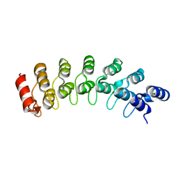
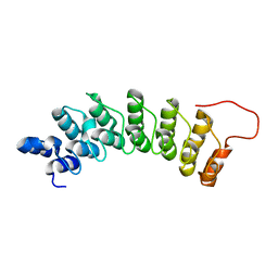
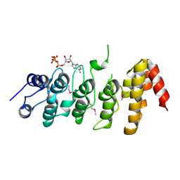
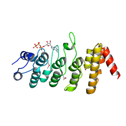
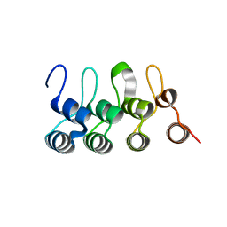
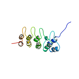 | | P19-INK4D FROM MOUSE, NMR, 20 STRUCTURES | | 分子名称: | P19-INK4D | | 著者 | Archer, S.J, Luh, F.Y, Domaille, P.J, Smith, B.O, Laue, E.D. | | 登録日 | 1997-07-25 | | 公開日 | 1998-09-16 | | 最終更新日 | 2022-02-16 | | 実験手法 | SOLUTION NMR | | 主引用文献 | Structure of the cyclin-dependent kinase inhibitor p19Ink4d.
Nature, 389, 1997
|
|
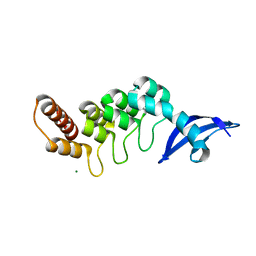
1BLX
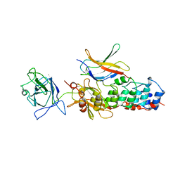
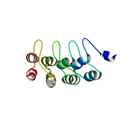
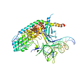
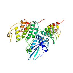 | | P19INK4D/CDK6 COMPLEX | | 分子名称: | CALCIUM ION, CYCLIN-DEPENDENT KINASE 6, P19INK4D | | 著者 | Brotherton, D.H, Dhanaraj, V, Wick, S, Brizuela, L, Domaille, P.J, Volyanik, E, Xu, X, Parisini, E, Smith, B.O, Archer, S.J, Serrano, M, Brenner, S.L, Blundell, T.L, Laue, E.D. | | 登録日 | 1998-07-21 | | 公開日 | 1999-06-01 | | 最終更新日 | 2023-08-02 | | 実験手法 | X-RAY DIFFRACTION (1.9 Å) | | 主引用文献 | Crystal structure of the complex of the cyclin D-dependent kinase Cdk6 bound to the cell-cycle inhibitor p19INK4d.
Nature, 395, 1998
|
|
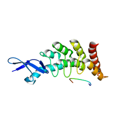
1D9S
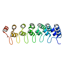
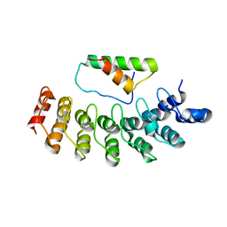
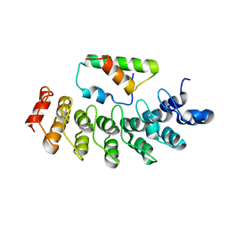
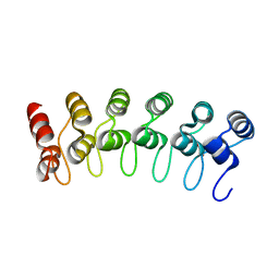
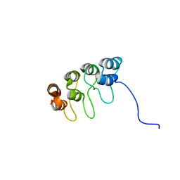 | | TUMOR SUPPRESSOR P15(INK4B) STRUCTURE BY COMPARATIVE MODELING AND NMR DATA | | 分子名称: | CYCLIN-DEPENDENT KINASE 4 INHIBITOR B | | 著者 | Yuan, C, Ji, L, Selby, T.L, Byeon, I.J.L, Tsai, M.D. | | 登録日 | 1999-10-29 | | 公開日 | 2000-07-28 | | 最終更新日 | 2022-02-16 | | 実験手法 | SOLUTION NMR | | 主引用文献 | Tumor suppressor INK4: comparisons of conformational properties between p16(INK4A) and p18(INK4C).
J.Mol.Biol., 294, 1999
|
|
1IKN
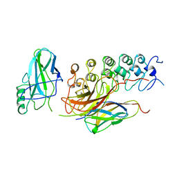 | | IKAPPABALPHA/NF-KAPPAB COMPLEX | | 分子名称: | PROTEIN (I-KAPPA-B-ALPHA), PROTEIN (NF-KAPPA-B P50D SUBUNIT), PROTEIN (NF-KAPPA-B P65 SUBUNIT) | | 著者 | Huxford, T, Huang, D.-B, Malek, S, Ghosh, G. | | 登録日 | 1998-11-13 | | 公開日 | 1999-04-12 | | 最終更新日 | 2023-08-16 | | 実験手法 | X-RAY DIFFRACTION (2.3 Å) | | 主引用文献 | The crystal structure of the IkappaBalpha/NF-kappaB complex reveals mechanisms of NF-kappaB inactivation.
Cell(Cambridge,Mass.), 95, 1998
|
|
1IXV
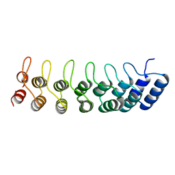
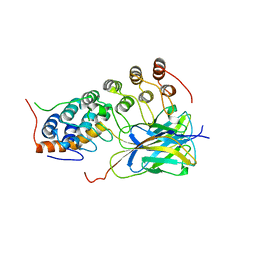
 | | Crystal Structure Analysis of homolog of oncoprotein gankyrin, an interactor of Rb and CDK4/6 | | 分子名称: | Probable 26S proteasome regulatory subunit p28 | | 著者 | Padmanabhan, B, Adachi, N, Kataoka, K, Horikoshi, M. | | 登録日 | 2002-07-09 | | 公開日 | 2003-12-16 | | 最終更新日 | 2023-12-27 | | 実験手法 | X-RAY DIFFRACTION (2.3 Å) | | 主引用文献 | Crystal structure of the homolog of the oncoprotein gankyrin, an interactor of Rb and CDK4/6
J.BIOL.CHEM., 279, 2004
|
|
1K1A
 | | Crystal structure of the ankyrin repeat domain of Bcl-3: a unique member of the IkappaB protein family | | 分子名称: | B-cell lymphoma 3-encoded protein | | 著者 | Michel, F, Soler-Lopez, M, Petosa, C, Cramer, P, Siebenlist, U, Mueller, C.W. | | 登録日 | 2001-09-24 | | 公開日 | 2001-11-21 | | 最終更新日 | 2023-08-16 | | 実験手法 | X-RAY DIFFRACTION (1.86 Å) | | 主引用文献 | Crystal structure of the ankyrin repeat domain of Bcl-3: a unique member of the IkappaB protein family.
EMBO J., 20, 2001
|
|
1K1B
 | | Crystal structure of the ankyrin repeat domain of Bcl-3: a unique member of the IkappaB protein family | | 分子名称: | B-cell lymphoma 3-encoded protein | | 著者 | Michel, F, Soler-Lopez, M, Petosa, C, Cramer, P, Siebenlist, U, Mueller, C.W. | | 登録日 | 2001-09-24 | | 公開日 | 2001-11-21 | | 最終更新日 | 2023-08-16 | | 実験手法 | X-RAY DIFFRACTION (1.9 Å) | | 主引用文献 | Crystal structure of the ankyrin repeat domain of Bcl-3: a unique member of the IkappaB protein family.
EMBO J., 20, 2001
|
|
1K3Z
 | | X-ray crystal structure of the IkBb/NF-kB p65 homodimer complex | | 分子名称: | Transcription factor p65, transcription factor inhibitor I-kappa-B-beta | | 著者 | Shiva, M, Huang, D.B, Chen, Y, Huxford, T, Ghosh, S, Ghosh, G. | | 登録日 | 2001-10-04 | | 公開日 | 2002-10-04 | | 最終更新日 | 2023-08-16 | | 実験手法 | X-RAY DIFFRACTION (2.5 Å) | | 主引用文献 | X-ray crystal structure of an IkappaBbeta x NF-kappaB p65 homodimer complex.
J.Biol.Chem., 278, 2003
|
|
1N11
 | | D34 REGION OF HUMAN ANKYRIN-R AND LINKER | | 分子名称: | Ankyrin, BROMIDE ION, CHLORIDE ION | | 著者 | Michaely, P, Tomchick, D.R, Machius, M, Anderson, R.G.W. | | 登録日 | 2002-10-16 | | 公開日 | 2002-12-11 | | 最終更新日 | 2024-02-14 | | 実験手法 | X-RAY DIFFRACTION (2.7 Å) | | 主引用文献 | Crystal structure of a 12 ANK repeat stack from human ankyrinR
Embo J., 21, 2002
|
|
1NFI
 | |
1OY3
 | | CRYSTAL STRUCTURE OF AN IKBBETA/NF-KB P65 HOMODIMER COMPLEX | | 分子名称: | Transcription factor p65, transcription factor inhibitor I-kappa-B-beta | | 著者 | Malek, S, Huang, D.B, Huxford, T, Ghosh, S, Ghosh, G. | | 登録日 | 2003-04-03 | | 公開日 | 2003-05-20 | | 最終更新日 | 2023-08-16 | | 実験手法 | X-RAY DIFFRACTION (2.05 Å) | | 主引用文献 | X-ray crystal structure of an IkappaBbeta x NF-kappaB p65 homodimer complex.
J.Biol.Chem., 278, 2003
|
|
1SW6
 | |
1WDY
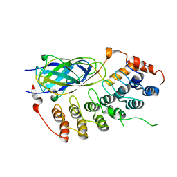
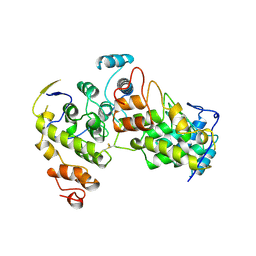
 | | Crystal structure of ribonuclease | | 分子名称: | 2-5A-dependent ribonuclease, 5'-O-MONOPHOSPHORYLADENYLYL(2'->5')ADENYLYL(2'->5')ADENOSINE | | 著者 | Tanaka, N, Nakanishi, M, Kusakabe, Y, Goto, Y, Kitade, Y, Nakamura, K.T. | | 登録日 | 2004-05-19 | | 公開日 | 2004-10-05 | | 最終更新日 | 2021-11-10 | | 実験手法 | X-RAY DIFFRACTION (1.8 Å) | | 主引用文献 | Structural basis for recognition of 2',5'-linked oligoadenylates by human ribonuclease L
Embo J., 23, 2004
|
|
1WG0
 | | Structural comparison of Nas6p protein structures in two different crystal forms | | 分子名称: | Probable 26S proteasome regulatory subunit p28 | | 著者 | Nakamura, Y, Umehara, T, Tanaka, A, Horikoshi, M, Yokoyama, S, Padmanabhan, B, RIKEN Structural Genomics/Proteomics Initiative (RSGI) | | 登録日 | 2004-05-27 | | 公開日 | 2005-06-07 | | 最終更新日 | 2023-10-25 | | 実験手法 | X-RAY DIFFRACTION (2.53 Å) | | 主引用文献 | Structural comparison of Nas6p protein structures in two different crystal forms
To be Published
|
|
1YCS
 | | P53-53BP2 COMPLEX | | 分子名称: | 53BP2, P53, ZINC ION | | 著者 | Gorina, S, Pavletich, N.P. | | 登録日 | 1996-09-30 | | 公開日 | 1997-11-19 | | 最終更新日 | 2024-02-14 | | 実験手法 | X-RAY DIFFRACTION (2.2 Å) | | 主引用文献 | Structure of the p53 tumor suppressor bound to the ankyrin and SH3 domains of 53BP2.
Science, 274, 1996
|
|
1YMP
 | |
2DZN
 | |
2DZO
 | |
2FO1
 | |
2NYJ
 | |
2PNN
 | |
2QC9
 | |
3C5R
 | |
3DEO
 | |
3DEP
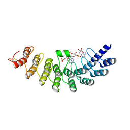
 | | Structural basis for specific substrate recognition by the chloroplast signal recognition particle protein cpSRP43 | | 分子名称: | CHLORIDE ION, Signal recognition particle 43 kDa protein, YPGGSFDPLGLA | | 著者 | Holdermann, I, Stengel, K.F, Wild, K, Sinning, I. | | 登録日 | 2008-06-10 | | 公開日 | 2008-08-12 | | 最終更新日 | 2023-11-01 | | 実験手法 | X-RAY DIFFRACTION (2.7 Å) | | 主引用文献 | Structural basis for specific substrate recognition by the chloroplast signal recognition particle protein cpSRP43.
Science, 321, 2008
|
|


