Loading
PDBj
MenuPDBj@FacebookPDBj@TwitterPDBj@YouTubewwPDB FoundationwwPDB
RCSB PDBPDBeBMRBAdv. SearchSearch help

7PVV
DownloadVisualize
BU of 7pvv by Molmil
Crystal structure of the Abl SH3 domain G92N-Y93N-N94T-H95E mutant
Descriptor: Bcr-abl1 e6a2 chimeric protein, DI(HYDROXYETHYL)ETHER, TRIETHYLENE GLYCOL
Authors:Camara-Artigas, A, Salinas Garcia, M.C.
Deposit date:2021-10-05
Release date:2022-09-14
Last modified:2024-01-31
Method:X-RAY DIFFRACTION (1.82 Å)
Cite:The effect of the hinge loops composition in the domain swapping of the SH3 domain
To be published
7PVT
DownloadVisualize
BU of 7pvt by Molmil
Crystal structure of the v-Src SH3 domain Q128R mutant in complex with the synthetic peptide VSL12
Descriptor: DI(HYDROXYETHYL)ETHER, GLYCEROL, Tyrosine-protein kinase transforming protein Src, ...
Authors:Camara-Artigas, A, Salinas-Garcia, M.C.
Deposit date:2021-10-05
Release date:2022-09-14
Last modified:2024-01-31
Method:X-RAY DIFFRACTION (1.6 Å)
Cite:Crystal structure of the v-Src SH3 domain Q128R mutant in complex with the synthetic peptide VSL12
To be published
3EG0
DownloadVisualize
BU of 3eg0 by Molmil
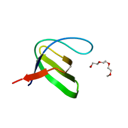
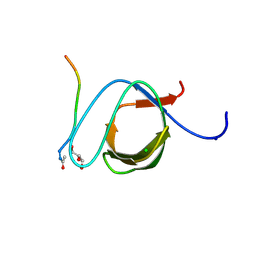
Crystal structure of the N114T mutant of ABL-SH3 domain
Descriptor: GLYCEROL, Proto-oncogene tyrosine-protein kinase ABL1
Authors:Camara-Artigas, A.
Deposit date:2008-09-10
Release date:2009-09-15
Last modified:2023-08-30
Method:X-RAY DIFFRACTION (2.3 Å)
Cite:Role of interfacial water molecules in proline-rich ligand recognition by the Src homology 3 domain of Abl.
J.Biol.Chem., 285, 2010
7PVZ
DownloadVisualize
BU of 7pvz by Molmil
Crystal structure of the intertwined dimer of the c-Src SH3 domain E93V-S94A-R95S-T96G mutant
Descriptor: Isoform 2 of Proto-oncogene tyrosine-protein kinase Src
Authors:Camara-Artigas, A, Salinas Garcia, M.C.
Deposit date:2021-10-05
Release date:2022-11-16
Last modified:2024-01-31
Method:X-RAY DIFFRACTION (2 Å)
Cite:The effect of the hinge loops composition in the domain swapping of the SH3 domain
To be published
3FJ5
DownloadVisualize
BU of 3fj5 by Molmil
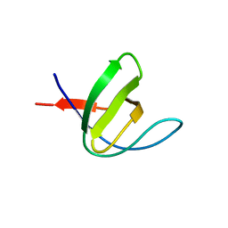
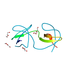
Crystal structure of the c-src-SH3 domain
Descriptor: ACETATE ION, GLYCEROL, Proto-oncogene tyrosine-protein kinase Src, ...
Authors:Camara-Artigas, A.
Deposit date:2008-12-14
Release date:2009-03-03
Last modified:2023-11-01
Method:X-RAY DIFFRACTION (1.65 Å)
Cite:Intertwined dimeric structure for the SH3 domain of the c-Src tyrosine kinase induced by polyethylene glycol binding
Febs Lett., 583, 2009
3M0P
DownloadVisualize
BU of 3m0p by Molmil
Crystal Structure of the R21D mutant of alpha-spectrin SH3 domain. Crystal obtained in ammonium sulphate at pH 4.
Descriptor: SULFATE ION, Spectrin alpha chain, brain
Authors:Camara-Artigas, A, Gavira, J.A.
Deposit date:2010-03-03
Release date:2011-01-19
Last modified:2023-09-06
Method:X-RAY DIFFRACTION (2.6 Å)
Cite:Understanding the polymorphic behaviour of a mutant of the alpha-spectrin SH3 domain by means of two 1.1 A structures
Acta Crystallogr.,Sect.D, 2011
3M0S
DownloadVisualize
BU of 3m0s by Molmil
Crystal Structure of the R21D mutant of alpha-spectrin SH3 domain. Crystal obtained in ammonium sulphate at pH 7
Descriptor: SULFATE ION, Spectrin alpha chain, brain
Authors:Camara-Artigas, A, Gavira, J.A.
Deposit date:2010-03-03
Release date:2011-01-19
Last modified:2023-09-06
Method:X-RAY DIFFRACTION (1.6 Å)
Cite:Understanding the polymorphic behaviour of a mutant of the alpha-spectrin SH3 domain by means of two 1.1 A structures
Acta Crystallogr.,Sect.D, 2011
3M0R
DownloadVisualize
BU of 3m0r by Molmil
Crystal Structure of the R21D mutant of alpha-spectrin SH3 domain. Crystal obtained in ammonium sulphate at pH 6.
Descriptor: SULFATE ION, Spectrin alpha chain, brain
Authors:Gavira, J.A, Camara-Artigas, A.
Deposit date:2010-03-03
Release date:2011-01-19
Last modified:2023-09-06
Method:X-RAY DIFFRACTION (1.1 Å)
Cite:Understanding the polymorphic behaviour of a mutant of the alpha-spectrin SH3 domain by means of two 1.1 A structures
Acta Crystallogr.,Sect.D, 2011
3M0T
DownloadVisualize
BU of 3m0t by Molmil
Crystal Structure of the R21D mutant of alpha-spectrin SH3 domain. Crystal obtained in ammonium sulphate at pH 9.
Descriptor: SULFATE ION, Spectrin alpha chain, brain
Authors:Camara-Artigas, A, Gavira, J.A.
Deposit date:2010-03-03
Release date:2011-01-19
Last modified:2024-02-21
Method:X-RAY DIFFRACTION (1.7 Å)
Cite:Understanding the polymorphic behaviour of a mutant of the alpha-spectrin SH3 domain by means of two 1.1 A structures
Acta Crystallogr.,Sect.D, 2011
3M0U
DownloadVisualize
BU of 3m0u by Molmil
Crystal Structure of the R21D mutant of alpha-spectrin SH3 domain. Hexagonal crystal obtained in sodium formate at pH 6.5.
Descriptor: FORMIC ACID, Spectrin alpha chain, brain
Authors:Gavira, J.A, Camara-Artigas, A.
Deposit date:2010-03-03
Release date:2011-01-19
Last modified:2023-09-06
Method:X-RAY DIFFRACTION (1.1 Å)
Cite:Understanding the polymorphic behaviour of a mutant of the alpha-spectrin SH3 domain by means of two 1.1 A structures
Acta Crystallogr.,Sect.D, 2011
3M0Q
DownloadVisualize
BU of 3m0q by Molmil
Crystal Structure of the R21D mutant of alpha-spectrin SH3 domain. Crystal obtained in ammonium sulphate at pH 5.
Descriptor: SULFATE ION, Spectrin alpha chain, brain
Authors:Camara-Artigas, A, Gavira, J.A.
Deposit date:2010-03-03
Release date:2011-01-19
Last modified:2023-09-06
Method:X-RAY DIFFRACTION (1.75 Å)
Cite:Understanding the polymorphic behaviour of a mutant of the alpha-spectrin SH3 domain by means of two 1.1 A structures
Acta Crystallogr.,Sect.D, 2011
3GBQ
DownloadVisualize
BU of 3gbq by Molmil
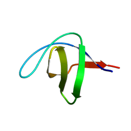
SOLUTION NMR STRUCTURE OF THE GRB2 N-TERMINAL SH3 DOMAIN COMPLEXED WITH A TEN-RESIDUE PEPTIDE DERIVED FROM SOS DIRECT REFINEMENT AGAINST NOES, J-COUPLINGS, AND 1H AND 13C CHEMICAL SHIFTS, MINIMIZED AVERAGE STRUCTURE
Descriptor: GRB2, SOS-1
Authors:Wittekind, M, Mapelli, C, Lee, V, Goldfarb, V, Friedrichs, M.S, Meyers, C.A, Mueller, L.
Deposit date:1996-12-23
Release date:1997-09-04
Last modified:2022-03-16
Method:SOLUTION NMR
Cite:Solution structure of the Grb2 N-terminal SH3 domain complexed with a ten-residue peptide derived from SOS: direct refinement against NOEs, J-couplings and 1H and 13C chemical shifts.
J.Mol.Biol., 267, 1997
3H0F
DownloadVisualize
BU of 3h0f by Molmil
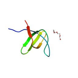
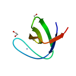
Crystal structure of the human Fyn SH3 R96W mutant
Descriptor: 1-METHOXY-2-[2-(2-METHOXY-ETHOXY]-ETHANE, Proto-oncogene tyrosine-protein kinase Fyn
Authors:Ponchon, L, Hoh, F, Labesse, G, Dumas, C, Arold, S.T.
Deposit date:2009-04-09
Release date:2010-04-21
Last modified:2023-11-01
Method:X-RAY DIFFRACTION (2.61 Å)
Cite:Synergy and allostery in ligand binding by HIV-1 Nef.
Biochem.J., 478, 2021
3H0I
DownloadVisualize
BU of 3h0i by Molmil
human Fyn SH3 domain R96I mutant, crystal form II
Descriptor: Proto-oncogene tyrosine-protein kinase Fyn
Authors:Dumas, C, Strub, M.-P, Arold, S.T.
Deposit date:2009-04-09
Release date:2010-04-21
Last modified:2023-11-01
Method:X-RAY DIFFRACTION (2.2 Å)
Cite:Synergy and allostery in ligand binding by HIV-1 Nef.
Biochem.J., 478, 2021
3H0H
DownloadVisualize
BU of 3h0h by Molmil
human Fyn SH3 domain R96I mutant, crystal form I
Descriptor: Proto-oncogene tyrosine-protein kinase Fyn, TETRAETHYLENE GLYCOL
Authors:Dumas, C, Strub, M.-P, Arold, S.T.
Deposit date:2009-04-09
Release date:2010-04-21
Last modified:2023-11-01
Method:X-RAY DIFFRACTION (1.76 Å)
Cite:Synergy and allostery in ligand binding by HIV-1 Nef.
Biochem.J., 478, 2021
3NGP
DownloadVisualize
BU of 3ngp by Molmil
High resolution structure of alpha-spectrin SH3 domain mutant with a redesigned core
Descriptor: Spectrin alpha chain, brain
Authors:Camara-Artigas, A, Gavira, J.A, Martin-Garcia, J.M.
Deposit date:2010-06-12
Release date:2010-09-08
Last modified:2023-09-06
Method:X-RAY DIFFRACTION (1.084 Å)
Cite:High-resolution structure of an alpha-spectrin SH3-domain mutant with a redesigned hydrophobic core.
Acta Crystallogr.,Sect.F, 66, 2010
3UA7
DownloadVisualize
BU of 3ua7 by Molmil
Crystal Structure of the Human Fyn SH3 domain in complex with a peptide from the Hepatitis C virus NS5A-protein
Descriptor: CHLORIDE ION, FORMIC ACID, GLYCEROL, ...
Authors:Martin-Garcia, J.M, Ruiz-Sanz, J, Luque, I, Camara-Artigas, A.
Deposit date:2011-10-21
Release date:2012-07-25
Last modified:2023-09-13
Method:X-RAY DIFFRACTION (1.5 Å)
Cite:The promiscuous binding of the Fyn SH3 domain to a peptide from the NS5A protein.
Acta Crystallogr.,Sect.D, 68, 2012
4OMO
DownloadVisualize
BU of 4omo by Molmil
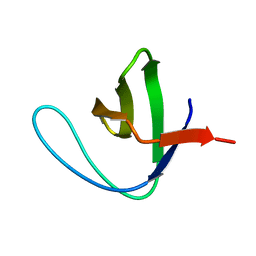
Crystal structure of the c-Src tyrosine kinase SH3 domain mutant Q128E
Descriptor: 4-(2-HYDROXYETHYL)-1-PIPERAZINE ETHANESULFONIC ACID, NICKEL (II) ION, Proto-oncogene tyrosine-protein kinase Src
Authors:Camara-Artigas, A, Bacarizo, J.
Deposit date:2014-01-27
Release date:2014-12-10
Last modified:2023-09-20
Method:X-RAY DIFFRACTION (1.04 Å)
Cite:Electrostatic Effects in the Folding of the SH3 Domain of the c-Src Tyrosine Kinase: pH-Dependence in 3D-Domain Swapping and Amyloid Formation.
Plos One, 9, 2014
4OML
DownloadVisualize
BU of 4oml by Molmil
Crystal structure of the intertwined dimer of the c-Src tyrosine kinase SH3 domain mutant Q128R
Descriptor: DI(HYDROXYETHYL)ETHER, Proto-oncogene tyrosine-protein kinase Src, TRIETHYLENE GLYCOL
Authors:Camara-Artigas, A, Bacarizo, J.
Deposit date:2014-01-27
Release date:2014-12-10
Last modified:2023-09-20
Method:X-RAY DIFFRACTION (1.6 Å)
Cite:Electrostatic Effects in the Folding of the SH3 Domain of the c-Src Tyrosine Kinase: pH-Dependence in 3D-Domain Swapping and Amyloid Formation.
Plos One, 9, 2014
4OMN
DownloadVisualize
BU of 4omn by Molmil
Crystal structure of the intertwined dimer of the c-Src tyrosine kinase SH3 domain mutant Q128E
Descriptor: DI(HYDROXYETHYL)ETHER, GLYCEROL, Proto-oncogene tyrosine-protein kinase Src, ...
Authors:Camara-Artigas, A, Bacarizo, J.
Deposit date:2014-01-27
Release date:2014-12-10
Last modified:2023-09-20
Method:X-RAY DIFFRACTION (1.5 Å)
Cite:Electrostatic Effects in the Folding of the SH3 Domain of the c-Src Tyrosine Kinase: pH-Dependence in 3D-Domain Swapping and Amyloid Formation.
Plos One, 9, 2014
4OMQ
DownloadVisualize
BU of 4omq by Molmil
Crystal structure of the intertwined dimer of the c-Src tyrosine kinase SH3 domain mutant S94A
Descriptor: DI(HYDROXYETHYL)ETHER, Proto-oncogene tyrosine-protein kinase Src, SULFATE ION, ...
Authors:Camara-Artigas, A, Bacarizo, J.
Deposit date:2014-01-27
Release date:2015-01-28
Last modified:2023-09-20
Method:X-RAY DIFFRACTION (2 Å)
Cite:The role of Gutamine 128 of the SH3 domain of the c-Src tyrosine kinase in 3D domain swapping and amyloid formation
To be Published
5FW9
DownloadVisualize
BU of 5fw9 by Molmil
Human Spectrin SH3 domain D48G, E7Y, K60Y
Descriptor: SPECTRIN ALPHA CHAIN, NON-ERYTHROCYTIC 1
Authors:Gallego, P, Navarro, S, Ventura, S, Reverter, D.
Deposit date:2016-02-12
Release date:2016-12-28
Last modified:2024-01-10
Method:X-RAY DIFFRACTION (1.55 Å)
Cite:Human Spectrin SH3 Domain D48G, E7Y, K60Y
To be Published
5FWC
DownloadVisualize
BU of 5fwc by Molmil
Human Spectrin SH3 domain D48G, E7A, K60A
Descriptor: 1,2-ETHANEDIOL, AMMONIUM ION, SPECTRIN ALPHA CHAIN, ...
Authors:Gallego, P, Navarro, S, Ventura, S, Reverter, D.
Deposit date:2016-02-12
Release date:2016-12-28
Last modified:2024-01-10
Method:X-RAY DIFFRACTION (1.4 Å)
Cite:Human Spectrin SH3 Domain D48G, E7A, K60A
To be Published
5FWB
DownloadVisualize
BU of 5fwb by Molmil
Human Spectrin SH3 domain D48G, E7F, K60F
Descriptor: SPECTRIN ALPHA CHAIN, NON-ERYTHROCYTIC 1
Authors:Gallego, P, Navarro, S, Ventura, S, Reverter, D.
Deposit date:2016-02-12
Release date:2016-12-28
Last modified:2024-05-08
Method:X-RAY DIFFRACTION (1.5 Å)
Cite:Human Spectrin SH3 Domain D48G, E7F, K60F
To be Published
4OMP
DownloadVisualize
BU of 4omp by Molmil
Crystal structure of the intertwined dimer of the c-Src tyrosine kinase SH3 domain mutant Q128K
Descriptor: DI(HYDROXYETHYL)ETHER, Proto-oncogene tyrosine-protein kinase Src, TRIETHYLENE GLYCOL
Authors:Camara-Artigas, A, Bacarizo, J.
Deposit date:2014-01-27
Release date:2014-12-10
Last modified:2023-09-20
Method:X-RAY DIFFRACTION (2 Å)
Cite:Electrostatic Effects in the Folding of the SH3 Domain of the c-Src Tyrosine Kinase: pH-Dependence in 3D-Domain Swapping and Amyloid Formation.
Plos One, 9, 2014

219869

건을2024-05-15부터공개중

PDB statisticsPDBj update infoContact PDBjnumon