Loading
PDBj
MenuPDBj@FacebookPDBj@TwitterPDBj@YouTubewwPDB FoundationwwPDB
RCSB PDBPDBeBMRBAdv. SearchSearch help

8W2L
DownloadVisualize
BU of 8w2l by Molmil
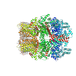
TRPM7 structure in complex with anticancer agent CCT128930 in closed state
Descriptor: (2S)-3-(hexadecanoyloxy)-2-[(9Z)-octadec-9-enoyloxy]propyl 2-(trimethylammonio)ethyl phosphate, 2-[2-[(1~{S},2~{S},4~{S},5'~{R},6~{R},7~{S},8~{R},9~{S},12~{S},13~{R},16~{S})-5',7,9,13-tetramethylspiro[5-oxapentacyclo[10.8.0.0^{2,9}.0^{4,8}.0^{13,18}]icos-18-ene-6,2'-oxane]-16-yl]oxyethyl]propane-1,3-diol, 4-(4-chlorobenzyl)-1-(7H-pyrrolo[2,3-d]pyrimidin-4-yl)piperidin-4-aminium, ...
Authors:Nadezhdin, K.D, Sobolevsky, A.I.
Deposit date:2024-02-20
Release date:2024-04-17
Last modified:2024-04-24
Method:ELECTRON MICROSCOPY (2.45 Å)
Cite:Structural basis of selective TRPM7 inhibition by the anticancer agent CCT128930.
Cell Rep, 43, 2024
8T6L
DownloadVisualize
BU of 8t6l by Molmil
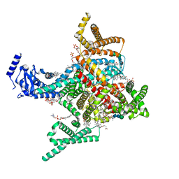
Cryo-EM structure of rat cardiac sodium channel NaV1.5 with batrachotoxin analog BTX-B
Descriptor: (1R)-1-[(5aR,7aR,9R,11aS,11bS,12R,13aR)-9,12-dihydroxy-2,11a-dimethyl-1,2,3,4,7a,8,9,10,11,11a,12,13-dodecahydro-7H-9,11b-epoxy-13a,5a-prop[1]enophenanthro[2,1-f][1,4]oxazepin-14-yl]ethyl benzoate, (3beta,14beta,17beta,25R)-3-[4-methoxy-3-(methoxymethyl)butoxy]spirost-5-en, 1-palmitoyl-2-oleoyl-sn-glycero-3-phosphocholine, ...
Authors:Tonggu, L, Wisedchaisri, G, Gamal El-Din, T.M, Zheng, N, Catterall, W.A.
Deposit date:2023-06-16
Release date:2024-03-06
Last modified:2024-03-27
Method:ELECTRON MICROSCOPY (3.3 Å)
Cite:Dual receptor-sites reveal the structural basis for hyperactivation of sodium channels by poison-dart toxin batrachotoxin.
Nat Commun, 15, 2024
4EN1
DownloadVisualize
BU of 4en1 by Molmil
The 1.62A structure of a FRET-optimized Cerulean Fluorescent Protein
Descriptor: ACETATE ION, DI(HYDROXYETHYL)ETHER, GLYCEROL, ...
Authors:Watkins, J.L.
Deposit date:2012-04-12
Release date:2013-04-24
Last modified:2023-12-06
Method:X-RAY DIFFRACTION (1.62 Å)
Cite:The 1.6 A resolution structure of a FRET-optimized Cerulean fluorescent protein.
Acta Crystallogr.,Sect.D, 69, 2013
6AA6
DownloadVisualize
BU of 6aa6 by Molmil
X-ray structure of ReQy1 (reduced form)
Descriptor: Green fluorescent protein
Authors:Kurisu, G, Yasuda, A, Tabushi, N, Tanaka, H.
Deposit date:2018-07-17
Release date:2019-05-29
Last modified:2023-11-22
Method:X-RAY DIFFRACTION (2.39 Å)
Cite:Multicolor redox sensor proteins can visualize redox changes in various compartments of the living cell.
Biochim Biophys Acta Gen Subj, 1863, 2019
6EFR
DownloadVisualize
BU of 6efr by Molmil
Crystal Structure of iNicSnFR 1.0
Descriptor: iNicSnFR 1.0, a genetically encoded nicotine biosensor,Green fluorescent protein
Authors:Shivange, A.V, Borden, P.M.
Deposit date:2018-08-17
Release date:2019-01-23
Last modified:2023-11-15
Method:X-RAY DIFFRACTION (2.4 Å)
Cite:Nicotinic Drugs in the Endoplasmic Reticulum: Beginning the Inside-out Pathway of Addiction and Therapy
J.Gen.Physiol., 2019
8A6O
DownloadVisualize
BU of 8a6o by Molmil
Room temperature rsEGFP2 with a chlorinated chromophore 600 fs after Photoexcitation
Descriptor: Green fluorescent protein
Authors:Fadini, A, van Thor, J.
Deposit date:2022-06-18
Release date:2023-07-19
Last modified:2024-02-07
Method:X-RAY DIFFRACTION (1.63 Å)
Cite:Serial Femtosecond Crystallography Reveals that Photoactivation in a Fluorescent Protein Proceeds via the Hula Twist Mechanism.
J.Am.Chem.Soc., 145, 2023
8A6P
DownloadVisualize
BU of 8a6p by Molmil
Room temperature rsEGFP2 with a chlorinated chromophore 900 fs after photoexcitation
Descriptor: Green fluorescent protein
Authors:Fadini, A, van Thor, J.
Deposit date:2022-06-18
Release date:2023-07-19
Last modified:2024-02-07
Method:X-RAY DIFFRACTION (1.63 Å)
Cite:Serial Femtosecond Crystallography Reveals that Photoactivation in a Fluorescent Protein Proceeds via the Hula Twist Mechanism.
J.Am.Chem.Soc., 145, 2023
8A6R
DownloadVisualize
BU of 8a6r by Molmil
Room temperature rsEGFP2 with a chlorinated chromophore 100 ps after Photoexcitation
Descriptor: Green fluorescent protein
Authors:Fadini, A, van Thor, J.
Deposit date:2022-06-18
Release date:2023-07-19
Last modified:2024-02-07
Method:X-RAY DIFFRACTION (1.63 Å)
Cite:Serial Femtosecond Crystallography Reveals that Photoactivation in a Fluorescent Protein Proceeds via the Hula Twist Mechanism.
J.Am.Chem.Soc., 145, 2023
8A6G
DownloadVisualize
BU of 8a6g by Molmil
Room temperature rsEGFP2 with a chlorinated chromophore in the non-fluorescent OFF-state
Descriptor: Green fluorescent protein
Authors:Fadini, A, van Thor, J.
Deposit date:2022-06-17
Release date:2023-07-19
Last modified:2024-02-07
Method:X-RAY DIFFRACTION (1.63 Å)
Cite:Serial Femtosecond Crystallography Reveals that Photoactivation in a Fluorescent Protein Proceeds via the Hula Twist Mechanism.
J.Am.Chem.Soc., 145, 2023
8A83
DownloadVisualize
BU of 8a83 by Molmil
rsEGFP2 with a chlorinated chromophore in the fluorescent ON-state in a crystal dehydrated after illumination
Descriptor: Green fluorescent protein, SULFATE ION
Authors:Fadini, A, van Thor, J, Chang, J.
Deposit date:2022-06-21
Release date:2023-07-19
Last modified:2024-02-07
Method:X-RAY DIFFRACTION (1.81 Å)
Cite:Serial Femtosecond Crystallography Reveals that Photoactivation in a Fluorescent Protein Proceeds via the Hula Twist Mechanism.
J.Am.Chem.Soc., 145, 2023
8A6N
DownloadVisualize
BU of 8a6n by Molmil
Room temperature rsEGFP2 with a chlorinated chromophore 300 fs after Photoexcitation
Descriptor: Green fluorescent protein
Authors:Fadini, A, van Thor, J.
Deposit date:2022-06-18
Release date:2023-07-19
Last modified:2024-02-07
Method:X-RAY DIFFRACTION (1.63 Å)
Cite:Serial Femtosecond Crystallography Reveals that Photoactivation in a Fluorescent Protein Proceeds via the Hula Twist Mechanism.
J.Am.Chem.Soc., 145, 2023
8A6Q
DownloadVisualize
BU of 8a6q by Molmil
Room temperature rsEGFP2 with a chlorinated chromophore 5 ps after photoexcitation
Descriptor: Green fluorescent protein
Authors:Fadini, A, van Thor, J.
Deposit date:2022-06-18
Release date:2023-07-19
Last modified:2024-02-07
Method:X-RAY DIFFRACTION (1.63 Å)
Cite:Serial Femtosecond Crystallography Reveals that Photoactivation in a Fluorescent Protein Proceeds via the Hula Twist Mechanism.
J.Am.Chem.Soc., 145, 2023
8A6S
DownloadVisualize
BU of 8a6s by Molmil
Room temperature rsEGFP2 with a chlorinated chromophore 1 microsecond after Photoexcitation
Descriptor: Green fluorescent protein
Authors:Fadini, A, van Thor, J.
Deposit date:2022-06-19
Release date:2023-07-19
Last modified:2024-02-07
Method:X-RAY DIFFRACTION (1.63 Å)
Cite:Serial Femtosecond Crystallography Reveals that Photoactivation in a Fluorescent Protein Proceeds via the Hula Twist Mechanism.
J.Am.Chem.Soc., 145, 2023
8A7V
DownloadVisualize
BU of 8a7v by Molmil
Room temperature rsEGFP2 in its OFF-state obtained with SFX
Descriptor: Green fluorescent protein
Authors:Fadini, A, van Thor, J.
Deposit date:2022-06-21
Release date:2023-07-19
Last modified:2024-02-07
Method:X-RAY DIFFRACTION (1.46 Å)
Cite:Serial Femtosecond Crystallography Reveals that Photoactivation in a Fluorescent Protein Proceeds via the Hula Twist Mechanism.
J.Am.Chem.Soc., 145, 2023
8AM4
DownloadVisualize
BU of 8am4 by Molmil
Cl-rsEGFP2 Long Wavelength Structure
Descriptor: Green fluorescent protein
Authors:Orr, C.M, Fadini, A, van Thor, J.
Deposit date:2022-08-02
Release date:2023-08-02
Last modified:2024-01-31
Method:X-RAY DIFFRACTION (2.02 Å)
Cite:Serial Femtosecond Crystallography Reveals that Photoactivation in a Fluorescent Protein Proceeds via the Hula Twist Mechanism.
J.Am.Chem.Soc., 2023
8BVG
DownloadVisualize
BU of 8bvg by Molmil
Bright fluorescent protein BrUSLEE with subnanosecond fluorescence lifetime
Descriptor: BrUSSLEE, GLYCEROL
Authors:Pletnev, V, Pletneva, N.
Deposit date:2022-12-03
Release date:2023-04-12
Last modified:2023-04-26
Method:X-RAY DIFFRACTION (2.38 Å)
Cite:Crystal Structure of Bright Fluorescent Protein BrUSLEE with Subnanosecond Fluorescence Lifetime; Electric and Dynamic Properties.
Int J Mol Sci, 24, 2023
8BXP
DownloadVisualize
BU of 8bxp by Molmil
SfGFP C148 F206 mutant
Descriptor: Green fluorescent protein
Authors:Ahmed, R.D, Rizkallah, P.J, Jones, D.D.
Deposit date:2022-12-09
Release date:2023-12-20
Method:X-RAY DIFFRACTION (1.79 Å)
Cite:SfGFP C148 F206 mutant
To Be Published
8C1X
DownloadVisualize
BU of 8c1x by Molmil
sfGFP C148 F206 mutant
Descriptor: Green fluorescent protein
Authors:Ahmed, R.D, Rizkallah, P.J, Jones, D.D.
Deposit date:2022-12-21
Release date:2024-01-10
Method:X-RAY DIFFRACTION (1.89 Å)
Cite:sfGFP C148 F206 mutant
To Be Published
4XBI
DownloadVisualize
BU of 4xbi by Molmil
Structure Of A Malarial Protein Involved in Proteostasis
Descriptor: ClpB protein, putative,Green fluorescent protein, SULFATE ION
Authors:Egea, P.F, Ah Young, A.P, Cascio, D.
Deposit date:2014-12-17
Release date:2015-07-29
Last modified:2023-11-15
Method:X-RAY DIFFRACTION (2.013 Å)
Cite:Structural mapping of the ClpB ATPases of Plasmodium falciparum: Targeting protein folding and secretion for antimalarial drug design.
Protein Sci., 24, 2015
6L27
DownloadVisualize
BU of 6l27 by Molmil
X-ray crystal structure of the mutant green fluorescent protein
Descriptor: Green fluorescent protein
Authors:Adachi, M, Shimizu, R, Shibazaki, C, Kagotani, Y, Ostermann, A, Schrader, T.E.
Deposit date:2019-10-02
Release date:2020-04-01
Last modified:2023-11-15
Method:X-RAY DIFFRACTION (0.77 Å)
Cite:Direct Observation of the Protonation States in the Mutant Green Fluorescent Protein.
J Phys Chem Lett, 11, 2020
5LEL
DownloadVisualize
BU of 5lel by Molmil
Crystal structure of DARPin-DARPin rigid fusion, variant DD_Off7_10_3G124 in complex with Maltose-binding Protein and Green Fluorescent Protein
Descriptor: DD_Off7_10_3G124, Green fluorescent protein, Maltose-binding periplasmic protein
Authors:Batyuk, A, Wu, Y, Mittl, P.R, Plueckthun, A.
Deposit date:2016-06-30
Release date:2017-11-15
Last modified:2019-10-16
Method:X-RAY DIFFRACTION (3.1 Å)
Cite:Rigidly connected multispecific artificial binders with adjustable geometries.
Sci Rep, 7, 2017
5LEM
DownloadVisualize
BU of 5lem by Molmil
Crystal structure of DARPin-DARPin rigid fusion, variant DD_Off7_11_3G124 in complex with Maltose-binding Protein and Green Fluorescent Protein
Descriptor: DD_Off7_11_3G124, Green fluorescent protein, Maltose-binding periplasmic protein
Authors:Batyuk, A, Wu, Y, Mittl, P.R, Plueckthun, A.
Deposit date:2016-06-30
Release date:2017-08-02
Last modified:2019-10-16
Method:X-RAY DIFFRACTION (2.98 Å)
Cite:Rigidly connected multispecific artificial binders with adjustable geometries.
Sci Rep, 7, 2017
6FLL
DownloadVisualize
BU of 6fll by Molmil
SPECTROSCOPIC AND STRUCTURAL STUDY OF QW, A EGFP MUTANT SHOWING PHOTOSWITCHING PROPERTIES
Descriptor: Green fluorescent protein
Authors:Garau, G, Margheritis, E.
Deposit date:2018-01-26
Release date:2019-02-06
Last modified:2024-01-17
Method:X-RAY DIFFRACTION (1.79 Å)
Cite:Role of Gln222 in Photoswitching of Aequorea Fluorescent Proteins: A Twisting and H-Bonding Affair?
Acs Chem.Biol., 13, 2018
1EML
DownloadVisualize
BU of 1eml by Molmil
GREEN FLUORESCENT PROTEIN FROM AEQUOREA VICTORIA, MUTANT
Descriptor: GREEN FLUORESCENT PROTEIN
Authors:Palm, G, Zdanov, A, Wlodawer, A.
Deposit date:1997-03-31
Release date:1997-08-20
Last modified:2023-11-15
Method:X-RAY DIFFRACTION (2.3 Å)
Cite:The structural basis for spectral variations in green fluorescent protein.
Nat.Struct.Biol., 4, 1997
1EMK
DownloadVisualize
BU of 1emk by Molmil
GREEN FLUORESCENT PROTEIN FROM AEQUOREA VICTORIA, MUTANT
Descriptor: GREEN FLUORESCENT PROTEIN
Authors:Palm, G, Zdanov, A, Wlodawer, A.
Deposit date:1997-03-31
Release date:1997-08-20
Last modified:2023-08-09
Method:X-RAY DIFFRACTION (2.1 Å)
Cite:The structural basis for spectral variations in green fluorescent protein.
Nat.Struct.Biol., 4, 1997

220472

건을2024-05-29부터공개중

PDB statisticsPDBj update infoContact PDBjnumon