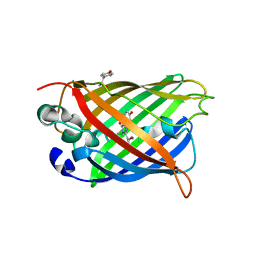
Loading
PDBj
MenuPDBj@FacebookPDBj@TwitterPDBj@YouTubewwPDB FoundationwwPDB
RCSB PDBPDBeBMRBAdv. SearchSearch help

8DHR
DownloadVisualize
BU of 8dhr by Molmil
An ester mutant of SfGFP
Descriptor: Green fluorescent protein
Authors:Reddi, R, Valiyaveetil, F.I.
Deposit date:2022-06-28
Release date:2024-01-17
Method:X-RAY DIFFRACTION (1.75 Å)
Cite:A facile approach for incorporating tyrosine esters to probe ion-binding sites and backbone hydrogen bonds.
J.Biol.Chem., 300, 2023
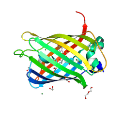
6AA2
DownloadVisualize
BU of 6aa2 by Molmil
X-ray structure of ReQy1 (oxidized form)
Descriptor: Green fluorescent protein
Authors:Sugiura, K, Yasuda, A, Tabushi, N, Tanaka, H, Kurisu, G, Hisabori, T.
Deposit date:2018-07-17
Release date:2019-05-29
Last modified:2023-11-22
Method:X-RAY DIFFRACTION (2.3 Å)
Cite:Multicolor redox sensor proteins can visualize redox changes in various compartments of the living cell.
Biochim Biophys Acta Gen Subj, 1863, 2019
8DPD
DownloadVisualize
BU of 8dpd by Molmil
superfolder GFP Tyr74pCNPhe mutant
Descriptor: 1,2-ETHANEDIOL, CARBON DIOXIDE, DI(HYDROXYETHYL)ETHER, ...
Authors:Phillips-Piro, C.M, Papoutsis, B, Piacentini, J.
Deposit date:2022-07-15
Release date:2022-08-31
Last modified:2023-11-15
Method:X-RAY DIFFRACTION (1.51 Å)
Cite:Superfolder GFP Tyr74pCNPhe mutant
To Be Published
6AA6
DownloadVisualize
BU of 6aa6 by Molmil
X-ray structure of ReQy1 (reduced form)
Descriptor: Green fluorescent protein
Authors:Kurisu, G, Yasuda, A, Tabushi, N, Tanaka, H.
Deposit date:2018-07-17
Release date:2019-05-29
Last modified:2023-11-22
Method:X-RAY DIFFRACTION (2.39 Å)
Cite:Multicolor redox sensor proteins can visualize redox changes in various compartments of the living cell.
Biochim Biophys Acta Gen Subj, 1863, 2019
6AA7
DownloadVisualize
BU of 6aa7 by Molmil
Fluorescent protein from Acropora digitifera
Descriptor: Fluorescent protein
Authors:Kim, S.E, Hwang, K.Y, Nam, K.H.
Deposit date:2018-07-17
Release date:2018-08-22
Last modified:2023-11-22
Method:X-RAY DIFFRACTION (1.8 Å)
Cite:Spectral and structural analysis of a red fluorescent protein from Acropora digitifera.
Protein Sci., 28, 2019
7Y40
DownloadVisualize
BU of 7y40 by Molmil
Crystal structure of a bright green fluorescent protein (StayGold) in jellyfish Cytaeis uchidae from Biortus
Descriptor: 1,2-ETHANEDIOL, staygold
Authors:Wu, J, Wang, F, Gui, W, Cheng, W, Yang, Y.
Deposit date:2022-06-13
Release date:2023-07-05
Last modified:2024-04-03
Method:X-RAY DIFFRACTION (1.7 Å)
Cite:Crystal structure of a bright green fluorescent protein (StayGold) in jellyfish Cytaeis uchidae from Biortus
To Be Published
2O2B
DownloadVisualize
BU of 2o2b by Molmil
Spectroscopic and Structural Study of the Heterotropic Linkage between Halide and Proton Ion Binding to Gfp Proteins: E2(GFP)-I Complex
Descriptor: Green fluorescent protein, IODIDE ION
Authors:Garau, G.
Deposit date:2006-11-29
Release date:2007-05-15
Last modified:2023-11-15
Method:X-RAY DIFFRACTION (1.94 Å)
Cite:Spectroscopic and Structural Study of Proton and Halide Ion Cooperative Binding to GFP.
Biophys.J., 93, 2007
8DTA
DownloadVisualize
BU of 8dta by Molmil
Metal sensitive GFP (mseGFP) complexed with phenylarsine oxide.
Descriptor: CALCIUM ION, DI(HYDROXYETHYL)ETHER, Green fluorescent protein, ...
Authors:Rosenbaum, J.C, Carlson, A.E.
Deposit date:2022-07-25
Release date:2023-07-05
Last modified:2024-01-31
Method:X-RAY DIFFRACTION (1.81 Å)
Cite:Glutathione modulates metal binding to proteins.
TBD
2OGR
DownloadVisualize
BU of 2ogr by Molmil
Crystal Structure of Yellow Fluorescent Protein from Zoanthus sp. at 1.8 A Resolution
Descriptor: FLUORESCENT PROTEIN FP538
Authors:Pletneva, N.V, Pletnev, S.V, Tikhonova, T.V, Pletnev, V.Z.
Deposit date:2007-01-08
Release date:2007-09-25
Last modified:2023-12-27
Method:X-RAY DIFFRACTION (1.8 Å)
Cite:Three-dimensional structure of yellow fluorescent protein zYFP538 from Zoanthus sp. at the resolution 1.8 angstrom
Bioorg.Khim., 33
7YDQ
DownloadVisualize
BU of 7ydq by Molmil
Structure of PfNT1(Y190A)-GFP in complex with GSK4
Descriptor: 5-methyl-N-[2-(2-oxidanylideneazepan-1-yl)ethyl]-2-phenyl-1,3-oxazole-4-carboxamide, Nucleoside transporter 1,Green fluorescent protein
Authors:Wang, C, Yu, L.Y, Li, J.L, Ren, R.B, Deng, D.
Deposit date:2022-07-04
Release date:2023-04-26
Last modified:2024-07-03
Method:ELECTRON MICROSCOPY (4.04 Å)
Cite:Structural basis of the substrate recognition and inhibition mechanism of Plasmodium falciparum nucleoside transporter PfENT1.
Nat Commun, 14, 2023
2OKW
DownloadVisualize
BU of 2okw by Molmil
A non-invasive GFP-based biosensor for mercury ions
Descriptor: Green fluorescent protein
Authors:Chapleau, R.R, Blomberg, R, Ford, P.C, Sagermann, M.
Deposit date:2007-01-17
Release date:2007-12-04
Last modified:2023-11-15
Method:X-RAY DIFFRACTION (1.9 Å)
Cite:Design of a highly specific and noninvasive biosensor suitable for real-time in vivo imaging of mercury (II) uptake.
Protein Sci., 17, 2008
6B9C
DownloadVisualize
BU of 6b9c by Molmil
Superfolder Green Fluorescent Protein with 4-nitro-L-phenylalanine at the chromophore (position 66)
Descriptor: CARBON DIOXIDE, Green fluorescent protein
Authors:Phillips-Piro, C.M, Brewer, S.H, Olenginski, G.M, Piacentini, J.
Deposit date:2017-10-10
Release date:2018-10-17
Last modified:2023-11-15
Method:X-RAY DIFFRACTION (1.695 Å)
Cite:Structural and spectrophotometric investigation of two unnatural amino-acid altered chromophores in the superfolder green fluorescent protein
Acta Crystallogr.,Sect.D, 2021
2OJK
DownloadVisualize
BU of 2ojk by Molmil
Crystal Structure of Green Fluorescent Protein from Zoanthus sp at 2.2 A Resolution
Descriptor: GFP-like fluorescent chromoprotein FP506
Authors:Pletneva, N.V, Pletnev, S.V, Tikhonova, T.V, Pletnev, V.Z.
Deposit date:2007-01-12
Release date:2007-09-25
Last modified:2023-11-15
Method:X-RAY DIFFRACTION (2.2 Å)
Cite:Refined crystal structures of red and green fluorescent proteins from the button polyp Zoanthus.
Acta Crystallogr.,Sect.D, 63, 2007
5Y8R
DownloadVisualize
BU of 5y8r by Molmil
ZsYellow at pH 3.5
Descriptor: GFP-like fluorescent chromoprotein FP538
Authors:Bae, J.E, Kim, I.J, Nam, K.H.
Deposit date:2017-08-21
Release date:2017-09-13
Last modified:2023-11-22
Method:X-RAY DIFFRACTION (2.3 Å)
Cite:Disruption of the hydrogen bonding network determines the pH-induced non-fluorescent state of the fluorescent protein ZsYellow by protonation of Glu221.
Biochem. Biophys. Res. Commun., 493, 2017
2O24
DownloadVisualize
BU of 2o24 by Molmil
Spectroscopic and Structural Study of the Heterotropic Linkage between Halide and Proton Ion Binding to Gfp Proteins: E2(GFP)-Cl Complex
Descriptor: CHLORIDE ION, Green fluorescent protein
Authors:Garau, G.
Deposit date:2006-11-29
Release date:2007-05-15
Last modified:2023-11-15
Method:X-RAY DIFFRACTION (1.45 Å)
Cite:Spectroscopic and Structural Study of Proton and Halide Ion Cooperative Binding to GFP.
Biophys.J., 93, 2007
2O29
DownloadVisualize
BU of 2o29 by Molmil
Spectroscopic and Structural Study of the Heterotropic Linkage between Halide and Proton Ion Binding to Gfp Proteins: E2(GFP)-BR Complex
Descriptor: BROMIDE ION, Green fluorescent protein
Authors:Garau, G.
Deposit date:2006-11-29
Release date:2007-05-15
Last modified:2023-11-15
Method:X-RAY DIFFRACTION (1.8 Å)
Cite:Spectroscopic and Structural Study of Proton and Halide Ion Cooperative Binding to GFP.
Biophys.J., 93, 2007
7YAO
DownloadVisualize
BU of 7yao by Molmil
Crystal structure of a bright green fluorescent protein (oxStayGold) in jellyfish Cytaeis uchidae from Biortus
Descriptor: oxstaygold
Authors:Wu, J, Wang, F, Gui, W, Cheng, W, Yang, Y.
Deposit date:2022-06-28
Release date:2023-07-19
Last modified:2024-04-03
Method:X-RAY DIFFRACTION (1.5 Å)
Cite:Crystal structure of a bright green fluorescent protein (oxStayGold) in jellyfish Cytaeis uchidae from Biortus
To Be Published
7YEU
DownloadVisualize
BU of 7yeu by Molmil
Superfolder green fluorescent protein with phosphine unnatural amino acid P3BF
Descriptor: Superfolder green fluorescent protein
Authors:Hu, C, Duan, H.Z, Liu, X.H, Chen, Y.X, Wang, J.Y.
Deposit date:2022-07-06
Release date:2023-07-26
Last modified:2024-02-28
Method:X-RAY DIFFRACTION (1.95 Å)
Cite:Genetically Encoded Phosphine Ligand for Metalloprotein Design.
J.Am.Chem.Soc., 144, 2022
2OKY
DownloadVisualize
BU of 2oky by Molmil
A non-invasive GFP-based biosensor for mercury ions
Descriptor: Green fluorescent protein
Authors:Sagermann, M, Chapleau, R.R.
Deposit date:2007-01-18
Release date:2007-12-04
Last modified:2023-11-15
Method:X-RAY DIFFRACTION (2.4 Å)
Cite:Design of a highly specific and noninvasive biosensor suitable for real-time in vivo imaging of mercury (II) uptake.
Protein Sci., 17, 2008
7Z7P
DownloadVisualize
BU of 7z7p by Molmil
Structure of the fluorescent protein NeonCyan0.95 at pH 5.6
Descriptor: NeonCyan0.95, SULFATE ION
Authors:Depernet, H, Dupuy, J, Royant, A.
Deposit date:2022-03-16
Release date:2022-05-04
Last modified:2024-01-31
Method:X-RAY DIFFRACTION (1.95 Å)
Cite:Cyan fluorescent proteins derived from mNeonGreen.
Protein Eng.Des.Sel., 35, 2022
7Z7O
DownloadVisualize
BU of 7z7o by Molmil
Structure of the fluorescent protein NeonCyan0.95 at pH 7.5
Descriptor: NeonCyan0.95
Authors:Clavel, D, Dupuy, J, Royant, A.
Deposit date:2022-03-16
Release date:2022-05-04
Last modified:2024-01-31
Method:X-RAY DIFFRACTION (1.51 Å)
Cite:Cyan fluorescent proteins derived from mNeonGreen.
Protein Eng.Des.Sel., 35, 2022
7Z7Q
DownloadVisualize
BU of 7z7q by Molmil
Structure of the T207D single-point mutant of the fluorescent protein NeonCyan1 at pH 6.5
Descriptor: NeonCyan1
Authors:Duarte, K, Dupuy, J, Royant, A.
Deposit date:2022-03-16
Release date:2022-05-04
Last modified:2024-01-31
Method:X-RAY DIFFRACTION (1.6 Å)
Cite:Cyan fluorescent proteins derived from mNeonGreen.
Protein Eng.Des.Sel., 35, 2022
2OTB
DownloadVisualize
BU of 2otb by Molmil
Crystal structure of a monomeric cyan fluorescent protein in the fluorescent state
Descriptor: GFP-like fluorescent chromoprotein cFP484
Authors:Henderson, J.N, Ai, H, Campbell, R.E, Remington, S.J.
Deposit date:2007-02-07
Release date:2007-04-03
Last modified:2023-11-15
Method:X-RAY DIFFRACTION (1.79 Å)
Cite:Structural basis for reversible photobleaching of a green fluorescent protein homologue.
Proc.Natl.Acad.Sci.Usa, 104, 2007
2P4M
DownloadVisualize
BU of 2p4m by Molmil
High pH structure of Rtms5 H146S variant
Descriptor: GFP-like non-fluorescent chromoprotein, IODIDE ION
Authors:Battad, J.M, Wilmann, P.G, Olsen, S, Byres, E, Smith, S.C, Dove, S.G, Turcic, K.N, Devenish, R.J, Rossjohn, J, Prescott, M.
Deposit date:2007-03-12
Release date:2007-04-03
Last modified:2023-11-15
Method:X-RAY DIFFRACTION (1.8 Å)
Cite:A structural basis for the pH-dependent increase in fluorescence efficiency of chromoproteins
J.Mol.Biol., 368, 2007
2OTE
DownloadVisualize
BU of 2ote by Molmil
Crystal structure of a monomeric cyan fluorescent protein in the photobleached state
Descriptor: ACETATE ION, GFP-like fluorescent chromoprotein cFP484
Authors:Henderson, J.N, Ai, H, Campbell, R.E, Remington, S.J.
Deposit date:2007-02-07
Release date:2007-04-03
Last modified:2023-11-15
Method:X-RAY DIFFRACTION (1.47 Å)
Cite:Structural basis for reversible photobleaching of a green fluorescent protein homologue.
Proc.Natl.Acad.Sci.Usa, 104, 2007

223532

PDB entries from 2024-08-07

PDB statisticsPDBj update infoContact PDBjnumon