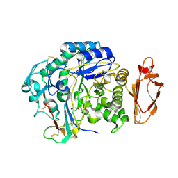
6WNU
 
 | |
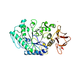
6WNI
 | |
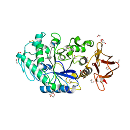
6XSJ
 | |
6XSV
 | |
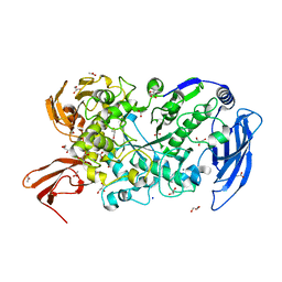
8CGT
 | | STRUCTURE OF CYCLODEXTRIN GLYCOSYLTRANSFERASE COMPLEXED WITH A THIO-MALTOHEXAOSE | | 分子名称: | CALCIUM ION, PROTEIN (CYCLODEXTRIN-GLYCOSYLTRANSFERASE), alpha-D-glucopyranose-(1-4)-4-thio-alpha-D-glucopyranose-(1-4)-alpha-D-glucopyranose-(1-4)-4-thio-alpha-D-glucopyranose-(1-4)-alpha-D-glucopyranose-(1-4)-4-thio-alpha-D-glucopyranose | | 著者 | Schmidt, A.K, Schulz, G.E. | | 登録日 | 1998-09-27 | | 公開日 | 1998-10-14 | | 最終更新日 | 2023-12-27 | | 実験手法 | X-RAY DIFFRACTION (2.4 Å) | | 主引用文献 | Substrate binding to a cyclodextrin glycosyltransferase and mutations increasing the gamma-cyclodextrin production.
Eur.J.Biochem., 255, 1998
|
|
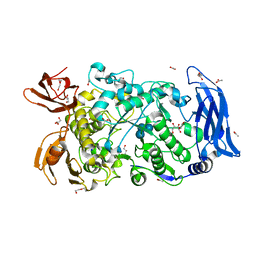
8CQG
 | | Crystal Structure of a Chimeric Alpha-Amylase from Pseudoalteromonas Haloplanktis | | 分子名称: | 1,2-ETHANEDIOL, Alpha-amylase, CALCIUM ION, ... | | 著者 | Skagseth, S, Lund, B.A, Griese, J.J, van der Ent, F, Aqvist, J. | | 登録日 | 2023-03-06 | | 公開日 | 2023-06-21 | | 最終更新日 | 2023-07-12 | | 実験手法 | X-RAY DIFFRACTION (1.74 Å) | | 主引用文献 | Computational design of the temperature optimum of an enzyme reaction.
Sci Adv, 9, 2023
|
|
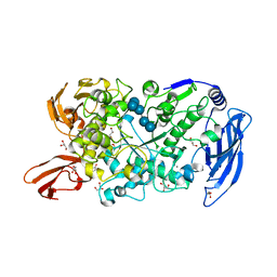
8CQF
 | | Crystal Structure of a Chimeric Alpha-Amylase from Pseudoalteromonas Haloplanktis Complexed with Rearranged Acarbose | | 分子名称: | 1,2-ETHANEDIOL, 4,6-dideoxy-4-{[(1S,4R,5S,6S)-4,5,6-trihydroxy-3-(hydroxymethyl)cyclohex-2-en-1-yl]amino}-alpha-D-glucopyranose-(1-4)-1,5-anhydro-D-glucitol, 4,6-dideoxy-4-{[(1S,4R,5S,6S)-4,5,6-trihydroxy-3-(hydroxymethyl)cyclohex-2-en-1-yl]amino}-alpha-D-glucopyranose-(1-4)-alpha-D-glucopyranose-(1-4)-alpha-D-glucopyranose, ... | | 著者 | Skagseth, S, Griese, J.J, Lund, B.A, van der Ent, F, Aqvist, J. | | 登録日 | 2023-03-06 | | 公開日 | 2023-06-21 | | 最終更新日 | 2023-07-12 | | 実験手法 | X-RAY DIFFRACTION (2.05 Å) | | 主引用文献 | Computational design of the temperature optimum of an enzyme reaction.
Sci Adv, 9, 2023
|
|
5JY7
 | |
5KEZ
 | |
7LST
 | | Ruminococcus bromii Amy12-D392A with 63-a-D-glucosyl-maltotriosyl-maltotriose | | 分子名称: | ACETATE ION, CALCIUM ION, GLYCEROL, ... | | 著者 | Koropatkin, N.M, Cockburn, D.W, Brown, H.A, Kibler, R.D. | | 登録日 | 2021-02-18 | | 公開日 | 2021-07-14 | | 最終更新日 | 2023-10-18 | | 実験手法 | X-RAY DIFFRACTION (2.05 Å) | | 主引用文献 | Structure and substrate recognition by the Ruminococcus bromii amylosome pullulanases.
J.Struct.Biol., 213, 2021
|
|
7LSU
 | | Ruminococcus bromii Amy12-D392A with 63-a-D-glucosyl-maltotriose | | 分子名称: | ACETATE ION, CALCIUM ION, DI(HYDROXYETHYL)ETHER, ... | | 著者 | Koropatkin, N.M, Cockburn, D.W, Brown, H.A, Kibler, R.D. | | 登録日 | 2021-02-18 | | 公開日 | 2021-07-14 | | 最終更新日 | 2023-10-18 | | 実験手法 | X-RAY DIFFRACTION (1.95 Å) | | 主引用文献 | Structure and substrate recognition by the Ruminococcus bromii amylosome pullulanases.
J.Struct.Biol., 213, 2021
|
|
7LSA
 | | Ruminococcus bromii Amy12 with maltoheptaose | | 分子名称: | CALCIUM ION, DI(HYDROXYETHYL)ETHER, GLYCEROL, ... | | 著者 | Koropatkin, N.M, Cockburn, D.W, Brown, H.A, Kibler, R.D. | | 登録日 | 2021-02-18 | | 公開日 | 2021-07-14 | | 最終更新日 | 2023-10-18 | | 実験手法 | X-RAY DIFFRACTION (1.76 Å) | | 主引用文献 | Structure and substrate recognition by the Ruminococcus bromii amylosome pullulanases.
J.Struct.Biol., 213, 2021
|
|
7LSR
 | | Ruminococcus bromii Amy12-D392A with maltoheptaose | | 分子名称: | CALCIUM ION, GLYCEROL, Pullulanase, ... | | 著者 | Koropatkin, N.M, Cockburn, D.W, Brown, H.A, Kibler, R.D. | | 登録日 | 2021-02-18 | | 公開日 | 2021-07-14 | | 最終更新日 | 2023-10-18 | | 実験手法 | X-RAY DIFFRACTION (2.42 Å) | | 主引用文献 | Structure and substrate recognition by the Ruminococcus bromii amylosome pullulanases.
J.Struct.Biol., 213, 2021
|
|
7LV6
 | | The structure of MalL mutant enzyme S536R from Bacillus subtilis | | 分子名称: | 2-AMINO-2-HYDROXYMETHYL-PROPANE-1,3-DIOL, CALCIUM ION, GLYCEROL, ... | | 著者 | Hamill, C.J, Prentice, E.J, Bahl, C.D, Truebridge, I.S, Arcus, V.L. | | 登録日 | 2021-02-24 | | 公開日 | 2022-03-09 | | 最終更新日 | 2023-10-18 | | 実験手法 | X-RAY DIFFRACTION (1.1 Å) | | 主引用文献 | Urea binding to guide rational design of mutations that influence enzyme dynamics
To Be Published
|
|
5MB2
 | |
5MAN
 | |
5M9X
 | | Structure of sucrose phosphorylase from Bifidobacterium adolescentis bound to glycosylated resveratrol | | 分子名称: | (2~{R},3~{S},4~{S},5~{R},6~{R})-2-(hydroxymethyl)-6-[3-[(~{E})-2-(4-hydroxyphenyl)ethenyl]-5-oxidanyl-phenoxy]oxane-3,4 ,5-triol, Sucrose phosphorylase | | 著者 | Grimm, C, Kraus, M. | | 登録日 | 2016-11-02 | | 公開日 | 2017-12-20 | | 最終更新日 | 2024-05-08 | | 実験手法 | X-RAY DIFFRACTION (2.349 Å) | | 主引用文献 | Switching enzyme specificity from phosphate to resveratrol glucosylation.
Chem. Commun. (Camb.), 53, 2017
|
|
7ML5
 | | Structure of the Starch Branching Enzyme I (BEI) complexed with maltododecaose from Oryza sativa L | | 分子名称: | Isoform 2 of 1,4-alpha-glucan-branching enzyme, chloroplastic/amyloplastic, alpha-D-glucopyranose-(1-4)-alpha-D-glucopyranose-(1-4)-alpha-D-glucopyranose-(1-4)-alpha-D-glucopyranose, ... | | 著者 | Nayebi Gavgani, H, Fawaz, R, Geiger, J.H. | | 登録日 | 2021-04-27 | | 公開日 | 2021-11-17 | | 最終更新日 | 2023-10-18 | | 実験手法 | X-RAY DIFFRACTION (2.35 Å) | | 主引用文献 | A structural explanation for the mechanism and specificity of plant branching enzymes I and IIb.
J.Biol.Chem., 298, 2021
|
|
8JJM
 | |
4WF7
 | | Crystal structures of trehalose synthase from Deinococcus radiodurans reveal that a closed conformation is involved in the intramolecular isomerization catalysis | | 分子名称: | 2-AMINO-2-HYDROXYMETHYL-PROPANE-1,3-DIOL, CALCIUM ION, MAGNESIUM ION, ... | | 著者 | Wang, Y.L, Chow, S.Y, Lin, Y.T, Hsieh, Y.C, Lee, G.C, Liaw, S.H. | | 登録日 | 2014-09-13 | | 公開日 | 2014-12-24 | | 最終更新日 | 2023-11-08 | | 実験手法 | X-RAY DIFFRACTION (2.21 Å) | | 主引用文献 | Structures of trehalose synthase from Deinococcus radiodurans reveal that a closed conformation is involved in catalysis of the intramolecular isomerization.
Acta Crystallogr.,Sect.D, 70, 2014
|
|
4WLC
 | | Structure of dextran glucosidase with glucose | | 分子名称: | CALCIUM ION, GLYCEROL, Glucan 1,6-alpha-glucosidase, ... | | 著者 | Kobayashi, M, Kato, K, Yao, M. | | 登録日 | 2014-10-07 | | 公開日 | 2015-08-26 | | 最終更新日 | 2023-11-08 | | 実験手法 | X-RAY DIFFRACTION (2.402 Å) | | 主引用文献 | Structural insights into the catalytic reaction that is involved in the reorientation of Trp238 at the substrate-binding site in GH13 dextran glucosidase
Febs Lett., 589, 2015
|
|
4X0N
 | |
8OR6
 | |
8ORP
 | | Crystal structure of Drosophila melanogaster alpha-amylase in complex with the inhibitor acarbose | | 分子名称: | 1,2-ETHANEDIOL, 4,6-dideoxy-4-{[(1S,2S,3S,4R,5R)-2,3,4-trihydroxy-5-(hydroxymethyl)cyclohexyl]amino}-alpha-D-glucopyranose-(1-4)-alpha-D-glucopyranose, 4,6-dideoxy-4-{[(1S,2S,3S,4R,5R)-2,3,4-trihydroxy-5-(hydroxymethyl)cyclohexyl]amino}-alpha-D-glucopyranose-(1-4)-alpha-D-glucopyranose-(1-4)-beta-D-glucopyranose, ... | | 著者 | Aghajari, N, Haser, R. | | 登録日 | 2023-04-16 | | 公開日 | 2023-08-16 | | 実験手法 | X-RAY DIFFRACTION (2 Å) | | 主引用文献 | Structural and Functional Characterization of Drosophila melanogaster alpha-Amylase.
Molecules, 28, 2023
|
|
4XB3
 | | Structure of dextran glucosidase | | 分子名称: | CALCIUM ION, Glucan 1,6-alpha-glucosidase, HEXAETHYLENE GLYCOL | | 著者 | Kobayashi, M, Kato, K, Yao, M. | | 登録日 | 2014-12-16 | | 公開日 | 2015-08-26 | | 最終更新日 | 2024-03-20 | | 実験手法 | X-RAY DIFFRACTION (2.093 Å) | | 主引用文献 | Structural insights into the catalytic reaction that is involved in the reorientation of Trp238 at the substrate-binding site in GH13 dextran glucosidase
Febs Lett., 589, 2015
|
|


