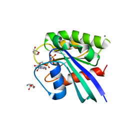
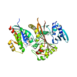
Loading
PDBj
MenuPDBj@FacebookPDBj@TwitterPDBj@YouTubewwPDB FoundationwwPDB
RCSB PDBPDBeBMRBAdv. SearchSearch help

4FMD
DownloadVisualize
BU of 4fmd by Molmil
EspG-Rab1 complex structure at 3.05 A
Descriptor: ALUMINUM FLUORIDE, DI(HYDROXYETHYL)ETHER, EspG protein, ...
Authors:Shao, F, Zhu, Y.
Deposit date:2012-06-16
Release date:2012-09-05
Last modified:2024-02-28
Method:X-RAY DIFFRACTION (3.05 Å)
Cite:Structurally Distinct Bacterial TBC-like GAPs Link Arf GTPase to Rab1 Inactivation to Counteract Host Defenses.
Cell(Cambridge,Mass.), 150, 2012
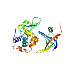
3E5T
DownloadVisualize
BU of 3e5t by Molmil
Crystal Structure Analysis of FP611
Descriptor: Red fluorescent protein eqFP611
Authors:Nar, H, Nienhaus, K, Nienhaus, U, Wiedenmann, J.
Deposit date:2008-08-14
Release date:2008-09-23
Last modified:2023-11-15
Method:X-RAY DIFFRACTION (1.1 Å)
Cite:Trans-cis isomerization is responsible for the red-shifted fluorescence in variants of the red fluorescent protein eqFP611.
J.Am.Chem.Soc., 130, 2008
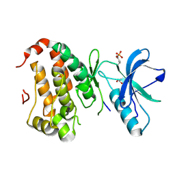
4GLA
DownloadVisualize
BU of 4gla by Molmil
OBody NL8 bound to hen egg-white lysozyme
Descriptor: Lysozyme C, OBody NL8
Authors:Steemson, J.D.
Deposit date:2012-08-14
Release date:2013-08-14
Last modified:2014-02-12
Method:X-RAY DIFFRACTION (2.75 Å)
Cite:Tracking Molecular Recognition at the Atomic Level with a New Protein Scaffold Based on the OB-Fold.
Plos One, 9, 2014
2QIH
DownloadVisualize
BU of 2qih by Molmil
Crystal structure of 527-665 fragment of UspA1 protein from Moraxella catarrhalis
Descriptor: CHLORIDE ION, PHOSPHATE ION, protein UspA1
Authors:Conners, R, Brady, R.L.
Deposit date:2007-07-04
Release date:2008-05-20
Last modified:2023-08-30
Method:X-RAY DIFFRACTION (1.897 Å)
Cite:The Moraxella adhesin UspA1 binds to its human CEACAM1 receptor by a deformable trimeric coiled-coil.
Embo J., 27, 2008
2QON
DownloadVisualize
BU of 2qon by Molmil
Human EphA3 kinase and juxtamembrane region, Y596F:Y602F:Y742A triple mutant
Descriptor: Ephrin receptor, GLYCEROL
Authors:Davis, T, Walker, J.R, Newman, E.M, Mackenzie, F, Butler-Cole, C, Weigelt, J, Sundstrom, M, Arrowsmith, C.H, Edwards, A.M, Bochkarev, A, Dhe-Paganon, S, Structural Genomics Consortium (SGC)
Deposit date:2007-07-20
Release date:2007-08-28
Last modified:2023-11-15
Method:X-RAY DIFFRACTION (1.79 Å)
Cite:Autoregulation by the Juxtamembrane Region of the Human Ephrin Receptor Tyrosine Kinase A3 (EphA3).
Structure, 16, 2008
3E3P
DownloadVisualize
BU of 3e3p by Molmil
Glycogen synthase kinase from Leishmania major
Descriptor: Protein kinase, putative Glycogen synthase kinase
Authors:Arakaki, T.L, Merritt, E.A.
Deposit date:2008-08-07
Release date:2008-08-19
Last modified:2024-02-21
Method:X-RAY DIFFRACTION (2 Å)
Cite:Glycogen synthase kinase from Leishmania major
To be Published
2HQ7
DownloadVisualize
BU of 2hq7 by Molmil
Crystal structure of Protein related to general stress protein 26(GS26) of B.subtilis (pyridoxinephosphate oxidase family) (NP_350077.1) from CLOSTRIDIUM ACETOBUTYLICUM at 2.00 A resolution
Descriptor: 1,2-ETHANEDIOL, CHLORIDE ION, Protein, ...
Authors:Joint Center for Structural Genomics (JCSG)
Deposit date:2006-07-18
Release date:2006-08-15
Last modified:2023-01-25
Method:X-RAY DIFFRACTION (2 Å)
Cite:Crystal structure of Protein related to general stress protein 26(GS26) of B.subtilis (pyridoxinephosphate oxidase family) (NP_350077.1) from CLOSTRIDIUM ACETOBUTYLICUM at 2.00 A resolution
To be published
3E7C
DownloadVisualize
BU of 3e7c by Molmil
Glucocorticoid Receptor LBD bound to GSK866
Descriptor: 5-amino-N-[(2S)-2-({[(2,6-dichlorophenyl)carbonyl](ethyl)amino}methyl)-3,3,3-trifluoro-2-hydroxypropyl]-1-(4-fluorophenyl)-1H-pyrazole-4-carboxamide, GLYCEROL, Glucocorticoid receptor, ...
Authors:Madauss, K.P, Williams, S.P, Mclay, I, Stewart, E.L, Bledsoe, R.K.
Deposit date:2008-08-18
Release date:2008-11-25
Last modified:2024-04-03
Method:X-RAY DIFFRACTION (2.15 Å)
Cite:The first X-ray crystal structure of the glucocorticoid receptor bound to a non-steroidal agonist.
Bioorg.Med.Chem.Lett., 18, 2008
3RO9
DownloadVisualize
BU of 3ro9 by Molmil
Candida glabrata dihydrofolate reductase complexed with NADPH and 6-ethyl-5-[(3R)-3-[3-methoxy-5-(pyridin-4-yl)phenyl]but-1-yn-1-yl]pyrimidine-2,4-diamine (UCP1006)
Descriptor: 6-ethyl-5-{(3R)-3-[3-methoxy-5-(pyridin-4-yl)phenyl]but-1-yn-1-yl}pyrimidine-2,4-diamine, NADPH DIHYDRO-NICOTINAMIDE-ADENINE-DINUCLEOTIDE PHOSPHATE, Strain CBS138 chromosome J complete sequence
Authors:Paulsen, J.L, Anderson, A.C.
Deposit date:2011-04-25
Release date:2012-05-09
Last modified:2024-02-28
Method:X-RAY DIFFRACTION (2.596 Å)
Cite:Structural analysis of the active sites of dihydrofolate reductase from two species of Candida uncovers ligand-induced conformational changes shared among species.
Bioorg.Med.Chem.Lett., 23, 2013
2QK0
DownloadVisualize
BU of 2qk0 by Molmil
Structural Basis of Microtubule Plus End Tracking by XMAP215, CLIP-170 and EB1
Descriptor: CAP-Gly domain-containing linker protein 1
Authors:Slep, K.C, Vale, R.D.
Deposit date:2007-07-09
Release date:2007-10-02
Last modified:2021-10-20
Method:X-RAY DIFFRACTION (2 Å)
Cite:Structural Basis of Microtubule Plus End Tracking by XMAP215, CLIP-170, and EB1.
Mol.Cell, 27, 2007
3E4B
DownloadVisualize
BU of 3e4b by Molmil
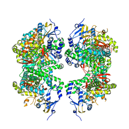
Crystal structure of AlgK from Pseudomonas fluorescens WCS374r
Descriptor: AlgK, CHLORIDE ION, GLYCEROL
Authors:Keiski, C.-L, Harwich, M, Jain, S, Neculai, A.M, Yip, P, Robinson, H, Whitney, J.C, Burrows, L.L, Ohman, D.E, Howell, P.L.
Deposit date:2008-08-11
Release date:2009-08-25
Last modified:2011-07-13
Method:X-RAY DIFFRACTION (2.5 Å)
Cite:AlgK is a TPR-containing protein and the periplasmic component of a novel exopolysaccharide secretin.
Structure, 18, 2010
3ROK
DownloadVisualize
BU of 3rok by Molmil
Crystal structure of human CD38 in complex with compound CZ-27
Descriptor: 5-O-[(S)-butoxy(hydroxy)phosphoryl]-2-deoxy-2-fluoro-alpha-D-arabinofuranose, ADP-ribosyl cyclase 1
Authors:Zhang, H, Lee, H.C, Hao, Q.
Deposit date:2011-04-26
Release date:2011-12-21
Last modified:2023-11-01
Method:X-RAY DIFFRACTION (1.65 Å)
Cite:Catalysis-based inhibitors of the calcium signaling function of CD38.
Biochemistry, 51, 2012
3E86
DownloadVisualize
BU of 3e86 by Molmil
High resolution Crystal Structure of the open NaK channel pore
Descriptor: (4S)-2-METHYL-2,4-PENTANEDIOL, CALCIUM ION, CESIUM ION, ...
Authors:Jiang, Y, Alam, A.
Deposit date:2008-08-19
Release date:2008-12-23
Last modified:2024-02-21
Method:X-RAY DIFFRACTION (1.6 Å)
Cite:High-resolution structure of the open NaK channel
Nat.Struct.Mol.Biol., 16, 2009
3E8F
DownloadVisualize
BU of 3e8f by Molmil
Crystal Structure of the the open NaK channel- K+/Ba2+
Descriptor: (4S)-2-METHYL-2,4-PENTANEDIOL, BARIUM ION, POTASSIUM ION, ...
Authors:Jiang, Y, Alam, A.
Deposit date:2008-08-19
Release date:2008-12-23
Last modified:2024-02-21
Method:X-RAY DIFFRACTION (2 Å)
Cite:Structural analysis of ion selectivity in the NaK channel
Nat.Struct.Mol.Biol., 16, 2009
3E7B
DownloadVisualize
BU of 3e7b by Molmil
Crystal Structure of Protein Phosphatase-1 Bound to the natural toxin inhibitor Tautomycin
Descriptor: (2Z)-2-[(1R)-3-{[(1R,2S,3R,6S,7S,10R)-10-{(2S,3S,6R,8S,9R)-3,9-dimethyl-8-[(3S)-3-methyl-4-oxopentyl]-1,7-dioxaspiro[5.5]undec-2-yl}-3,7-dihydroxy-2-methoxy-6-methyl-1-(1-methylethyl)-5-oxoundecyl]oxy}-1-hydroxy-3-oxopropyl]-3-methylbut-2-enedioic acid, AZIDE ION, CHLORIDE ION, ...
Authors:Kelker, M.S, Page, R, Peti, W.
Deposit date:2008-08-18
Release date:2008-11-04
Last modified:2023-08-30
Method:X-RAY DIFFRACTION (1.7 Å)
Cite:Crystal structures of protein phosphatase-1 bound to nodularin-R and tautomycin: a novel scaffold for structure-based drug design of serine/threonine phosphatase inhibitors
J.Mol.Biol., 385, 2009
2QLP
DownloadVisualize
BU of 2qlp by Molmil
Bifunctional dCTP deaminase:dUTPase from Mycobacterium tuberculosis, apo form
Descriptor: Deoxycytidine triphosphate deaminase, PENTAETHYLENE GLYCOL
Authors:Christophersen, S, Harris, P, Willemoes, M.
Deposit date:2007-07-13
Release date:2008-02-19
Last modified:2024-02-21
Method:X-RAY DIFFRACTION (2.47 Å)
Cite:Mechanism of dTTP inhibition of the bifunctional dCTP deaminase:dUTPase encoded by Mycobacterium tuberculosis
J.Mol.Biol., 376, 2007
4FYJ
DownloadVisualize
BU of 4fyj by Molmil
Crystal Structure of P. aeruginosa peptidyl-tRNA hydrolase
Descriptor: Peptidyl-tRNA hydrolase
Authors:Hughes, R.C, McFeeters, H, McFeeters, R.L.
Deposit date:2012-07-04
Release date:2012-12-12
Last modified:2023-09-13
Method:X-RAY DIFFRACTION (1.77 Å)
Cite:Recombinant production, crystallization and X-ray crystallographic structure determination of the peptidyl-tRNA hydrolase of Pseudomonas aeruginosa.
Acta Crystallogr.,Sect.F, 68, 2012
2QOD
DownloadVisualize
BU of 2qod by Molmil
Human EphA3 kinase and juxtamembrane region, Y602F mutant
Descriptor: Ephrin receptor
Authors:Davis, T, Walker, J.R, Newman, E.M, Mackenzie, F, Butler-Cole, C, Weigelt, J, Sundstrom, M, Arrowsmith, C.H, Edwards, A.M, Bochkarev, A, Dhe-Paganon, S, Structural Genomics Consortium (SGC)
Deposit date:2007-07-20
Release date:2007-08-21
Last modified:2023-08-30
Method:X-RAY DIFFRACTION (1.15 Å)
Cite:Autoregulation by the Juxtamembrane Region of the Human Ephrin Receptor Tyrosine Kinase A3 (EphA3).
Structure, 16, 2008
3RSO
DownloadVisualize
BU of 3rso by Molmil
H-Ras soaked in 20% S,R,S-bisfuranol: 1 of 10 in MSCS set
Descriptor: (3S,3aR,6aS)-hexahydrofuro[2,3-b]furan-3-ol, CALCIUM ION, GTPase HRas, ...
Authors:Mattos, C, Buhrman, G, Kearney, B.
Deposit date:2011-05-02
Release date:2011-09-21
Last modified:2024-02-28
Method:X-RAY DIFFRACTION (1.6 Å)
Cite:Analysis of Binding Site Hot Spots on the Surface of Ras GTPase.
J.Mol.Biol., 413, 2011
4FDH
DownloadVisualize
BU of 4fdh by Molmil
Structure of human aldosterone synthase, CYP11B2, in complex with fadrozole
Descriptor: 4-[(5R)-5,6,7,8-tetrahydroimidazo[1,5-a]pyridin-5-yl]benzonitrile, Cytochrome P450 11B2, mitochondrial, ...
Authors:Strushkevich, N, Shen, L, Tempel, W, Arrowsmith, C, Edwards, A, Usanov, S.A, Park, H.-W.
Deposit date:2012-05-28
Release date:2013-01-30
Last modified:2024-02-28
Method:X-RAY DIFFRACTION (2.71 Å)
Cite:Structural insights into aldosterone synthase substrate specificity and targeted inhibition.
Mol.Endocrinol., 27, 2013
2QV8
DownloadVisualize
BU of 2qv8 by Molmil
Structure of the minor pseudopilin EpsH from the Type 2 Secretion System of Vibrio cholerae
Descriptor: General secretion pathway protein H
Authors:Yanez, M.E, Korotkov, K.V, Abendroth, J, Hol, W.G.J.
Deposit date:2007-08-07
Release date:2008-02-26
Last modified:2017-10-25
Method:X-RAY DIFFRACTION (2 Å)
Cite:Structure of the minor pseudopilin EpsH from the Type 2 secretion system of Vibrio cholerae.
J.Mol.Biol., 377, 2008
2QVI
DownloadVisualize
BU of 2qvi by Molmil
Crystal structure of N-cadherin domains EC12
Descriptor: CALCIUM ION, Cadherin-2
Authors:Shapiro, L.S, Carroll, K.J.
Deposit date:2007-08-08
Release date:2008-08-12
Last modified:2024-02-21
Method:X-RAY DIFFRACTION (3.012 Å)
Cite:Biophysical characterization of N-cadherin and E-cadherin homophilic and heterophilic interactions
To be Published
2QPL
DownloadVisualize
BU of 2qpl by Molmil
Crystal structure of calf spleen purine nucleoside phosphorylase complexed to a novel purine analogue
Descriptor: 4-amino-7-methylpyrazolo[1,5-a][1,3,5]triazin-2(1H)-one, MAGNESIUM ION, Purine nucleoside phosphorylase, ...
Authors:Pereira, H.M, Berdini, V, Cleasby, A, Garratt, R.C.
Deposit date:2007-07-24
Release date:2007-12-18
Last modified:2023-11-29
Method:X-RAY DIFFRACTION (2.1 Å)
Cite:Crystal structure of calf spleen purine nucleoside phosphorylase complexed to a novel purine analogue.
Febs Lett., 581, 2007
2QQK
DownloadVisualize
BU of 2qqk by Molmil
Neuropilin-2 a1a2b1b2 Domains in Complex with a Semaphorin-Blocking Fab
Descriptor: 2-acetamido-2-deoxy-beta-D-glucopyranose, Antibody Heavy Chain, Antibody Light Chain, ...
Authors:Appleton, B.A, Wiesmann, C.
Deposit date:2007-07-26
Release date:2007-11-20
Last modified:2023-08-30
Method:X-RAY DIFFRACTION (2.75 Å)
Cite:Structural studies of neuropilin/antibody complexes provide insights into semaphorin and VEGF binding
Embo J., 26, 2007
3R1J
DownloadVisualize
BU of 3r1j by Molmil
Crystal structure of Alpha-ketoglutarate-dependent taurine dioxygenase from Mycobacterium avium, native form
Descriptor: 1,2-ETHANEDIOL, Alpha-ketoglutarate-dependent taurine dioxygenase, CHLORIDE ION, ...
Authors:Seattle Structural Genomics Center for Infectious Disease (SSGCID)
Deposit date:2011-03-10
Release date:2011-04-06
Last modified:2023-09-13
Method:X-RAY DIFFRACTION (2.05 Å)
Cite:Increasing the structural coverage of tuberculosis drug targets.
Tuberculosis (Edinb), 95, 2015

223790

PDB entries from 2024-08-14

PDB statisticsPDBj update infoContact PDBjnumon