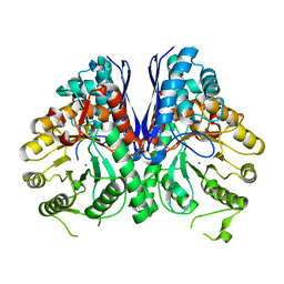
6D3Q
 
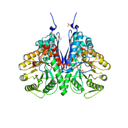
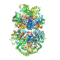
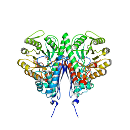 | | Crystal structure of Escherichia coli enolase complexed with a natural inhibitor SF2312. | | 分子名称: | Enolase, GLYCEROL, MAGNESIUM ION, ... | | 著者 | Erlandsen, H, Krucinska, J, Hazeen, A, Wright, D. | | 登録日 | 2018-04-16 | | 公開日 | 2019-11-27 | | 最終更新日 | 2023-10-04 | | 実験手法 | X-RAY DIFFRACTION (2.24 Å) | | 主引用文献 | Functional and structural basis of E. coli enolase inhibition by SF2312: a mimic of the carbanion intermediate.
Sci Rep, 9, 2019
|
|
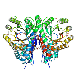
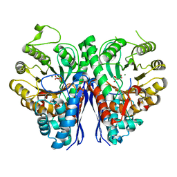
6ENL
 | |
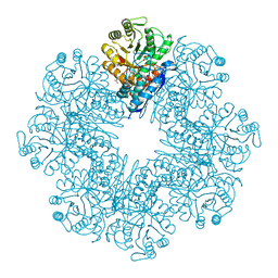
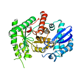
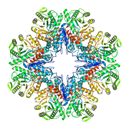
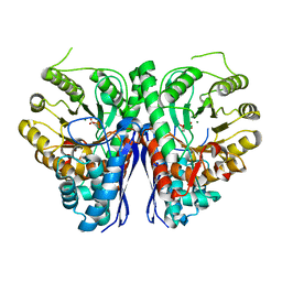
6J36
 | | crystal structure of Mycoplasma hyopneumoniae Enolase | | 分子名称: | Enolase, GLYCEROL, SULFATE ION | | 著者 | Chen, R, Zhang, S, Gan, R, Xie, X, Feng, Z, Wang, W, Ran, T, Zhang, W, Xiang, Q, Shao, G. | | 登録日 | 2019-01-04 | | 公開日 | 2019-05-15 | | 最終更新日 | 2023-11-22 | | 実験手法 | X-RAY DIFFRACTION (2.301 Å) | | 主引用文献 | Featured Species-Specific Loops Are Found in the Crystal Structure ofMhpEno, a Cell Surface Adhesin FromMycoplasma hyopneumoniae.
Front Cell Infect Microbiol, 9, 2019
|
|
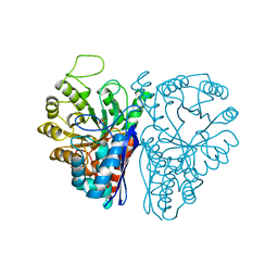
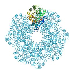
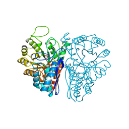
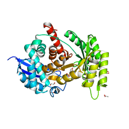
6L7D
 | | Mycobacterium tuberculosis enolase mutant - S42A | | 分子名称: | 1,2-ETHANEDIOL, 2-PHOSPHOGLYCERIC ACID, ACETATE ION, ... | | 著者 | Ahmad, M, Jha, B, Tiwari, S, Dwivedy, A, Biswal, B.K. | | 登録日 | 2019-11-01 | | 公開日 | 2020-11-04 | | 最終更新日 | 2023-11-22 | | 実験手法 | X-RAY DIFFRACTION (3 Å) | | 主引用文献 | Structural snapshots of Mycobacterium tuberculosis enolase reveal dual mode of 2PG binding and its implication in enzyme catalysis.
Iucrj, 10, 2023
|
|
6NB2
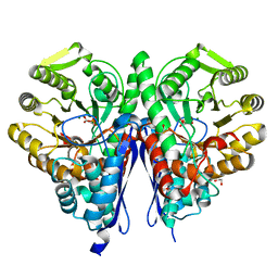 | |
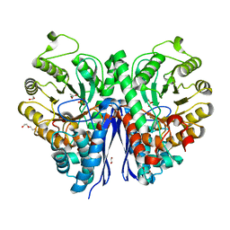
6NPF
 | | Structure of E.coli enolase in complex with an analog of the natural product SF-2312 metabolite. | | 分子名称: | Enolase, GLYCEROL, L(+)-TARTARIC ACID, ... | | 著者 | Erlandsen, H, Krucinska, J, Lombardo, M, Wright, D. | | 登録日 | 2019-01-17 | | 公開日 | 2019-11-27 | | 最終更新日 | 2023-10-11 | | 実験手法 | X-RAY DIFFRACTION (2.57 Å) | | 主引用文献 | Functional and structural basis of E. coli enolase inhibition by SF2312: a mimic of the carbanion intermediate.
Sci Rep, 9, 2019
|
|
6O4N
 | |
7CKP
 | | Mycobacterium tuberculosis Enolase | | 分子名称: | (4S)-2-METHYL-2,4-PENTANEDIOL, Enolase, MAGNESIUM ION | | 著者 | Biswal, B.K, Ahmad, M, Jha, B. | | 登録日 | 2020-07-18 | | 公開日 | 2021-07-21 | | 最終更新日 | 2023-11-29 | | 実験手法 | X-RAY DIFFRACTION (2.9 Å) | | 主引用文献 | Structural snapshots of Mycobacterium tuberculosis enolase reveal dual mode of 2PG binding and its implication in enzyme catalysis.
Iucrj, 10, 2023
|
|
7CLK
 | | Mycobacterium tuberculosis enolase in complex with alternate 2-phosphoglycerate | | 分子名称: | 1,2-ETHANEDIOL, 2-PHOSPHOGLYCERIC ACID, ACETATE ION, ... | | 著者 | Ahmad, M, Jha, B, Tiwari, S, Pal, R.K, Biswal, B.K. | | 登録日 | 2020-07-21 | | 公開日 | 2022-01-26 | | 最終更新日 | 2023-11-29 | | 実験手法 | X-RAY DIFFRACTION (2.15 Å) | | 主引用文献 | Structural snapshots of Mycobacterium tuberculosis enolase reveal dual mode of 2PG binding and its implication in enzyme catalysis.
Iucrj, 10, 2023
|
|
7CLL
 | | Mycobacterium tubeculosis enolase in complex with 2-Phosphoglycerate | | 分子名称: | 2-PHOSPHOGLYCERIC ACID, ACETATE ION, CHLORIDE ION, ... | | 著者 | Ahmad, M, Jha, B, Tiwari, S, Pal, R.K, Biswal, B.K. | | 登録日 | 2020-07-21 | | 公開日 | 2021-07-28 | | 最終更新日 | 2023-11-29 | | 実験手法 | X-RAY DIFFRACTION (1.99 Å) | | 主引用文献 | Structural snapshots of Mycobacterium tuberculosis enolase reveal dual mode of 2PG binding and its implication in enzyme catalysis.
Iucrj, 10, 2023
|
|
7DLR
 | | Mycobacterium tuberculosis enolase mutant - E163A | | 分子名称: | 1,2-ETHANEDIOL, ACETATE ION, CHLORIDE ION, ... | | 著者 | Ahmad, M, Biswal, B.K. | | 登録日 | 2020-11-30 | | 公開日 | 2021-12-01 | | 最終更新日 | 2023-11-29 | | 実験手法 | X-RAY DIFFRACTION (2.25 Å) | | 主引用文献 | Structural snapshots of Mycobacterium tuberculosis enolase reveal dual mode of 2PG binding and its implication in enzyme catalysis.
Iucrj, 10, 2023
|
|
7E2P
 | | The Crystal Structure of Mycoplasma bovis enolase | | 分子名称: | Enolase | | 著者 | Chen, R, Zhang, S, Gan, R, Wang, W, Ran, T, Shao, G, Xiong, Q, Feng, Z. | | 登録日 | 2021-02-07 | | 公開日 | 2022-02-02 | | 最終更新日 | 2023-11-29 | | 実験手法 | X-RAY DIFFRACTION (1.7 Å) | | 主引用文献 | Evidence for the Rapid and Divergent Evolution of Mycoplasmas: Structural and Phylogenetic Analysis of Enolases.
Front Mol Biosci, 8, 2022
|
|
7E2Q
 | | Crystal structure of Mycoplasma pneumoniae Enolase | | 分子名称: | Enolase, SULFATE ION | | 著者 | Chen, R, Zhang, S, Gan, R, Wang, W, Ran, T, Xiong, Q, Shao, G, Feng, Z. | | 登録日 | 2021-02-07 | | 公開日 | 2022-02-02 | | 最終更新日 | 2023-11-29 | | 実験手法 | X-RAY DIFFRACTION (1.8 Å) | | 主引用文献 | Evidence for the Rapid and Divergent Evolution of Mycoplasmas: Structural and Phylogenetic Analysis of Enolases.
Front Mol Biosci, 8, 2022
|
|
7E4F
 | | Mycobacterium tuberculosis enolase mutant - E204A complex with phosphoenolpyruvate | | 分子名称: | 1,2-ETHANEDIOL, ACETATE ION, DI(HYDROXYETHYL)ETHER, ... | | 著者 | Ahmad, M, Pal, R.K, Biswal, B.K. | | 登録日 | 2021-02-11 | | 公開日 | 2022-02-16 | | 最終更新日 | 2023-11-29 | | 実験手法 | X-RAY DIFFRACTION (2.3 Å) | | 主引用文献 | Structural snapshots of Mycobacterium tuberculosis enolase reveal dual mode of 2PG binding and its implication in enzyme catalysis.
Iucrj, 10, 2023
|
|
7E4X
 | |
7E51
 | |
7ENL
 | |
7MBH
 | | Structure of Human Enolase 2 in complex with phosphoserine | | 分子名称: | 1,2-ETHANEDIOL, ACETATE ION, Gamma-enolase, ... | | 著者 | Leonard, P.G, Hicks, K.G, Rutter, J. | | 登録日 | 2021-03-31 | | 公開日 | 2022-11-09 | | 最終更新日 | 2023-10-25 | | 実験手法 | X-RAY DIFFRACTION (2.1 Å) | | 主引用文献 | Protein-metabolite interactomics of carbohydrate metabolism reveal regulation of lactate dehydrogenase.
Science, 379, 2023
|
|
7N6G
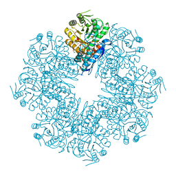
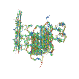 | | C1 of central pair | | 分子名称: | CPC1, Calmodulin, DPY30, ... | | 著者 | Han, L, Zhang, K. | | 登録日 | 2021-06-08 | | 公開日 | 2022-05-18 | | 最終更新日 | 2022-06-01 | | 実験手法 | ELECTRON MICROSCOPY (3.6 Å) | | 主引用文献 | Cryo-EM structure of an active central apparatus.
Nat.Struct.Mol.Biol., 29, 2022
|
|
7RHV
 | | Aspergillus fumigatus Enolase | | 分子名称: | Enolase, MAGNESIUM ION | | 著者 | Nguyen, S, Bruning, J.B. | | 登録日 | 2021-07-19 | | 公開日 | 2022-03-16 | | 最終更新日 | 2023-10-18 | | 実験手法 | X-RAY DIFFRACTION (2 Å) | | 主引用文献 | A structural model of the human plasminogen and Aspergillus fumigatus enolase complex.
Proteins, 90, 2022
|
|
7RHW
 | |
7RI0
 | |
7SQC
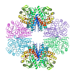
 | | Ciliary C1 central pair apparatus isolated from Chlamydomonas reinhardtii | | 分子名称: | ADENOSINE-5'-DIPHOSPHATE, CPC1, Calmodulin, ... | | 著者 | Gui, M, Wang, X, Dutcher, S.K, Brown, A, Zhang, R. | | 登録日 | 2021-11-05 | | 公開日 | 2022-04-13 | | 最終更新日 | 2022-06-08 | | 実験手法 | ELECTRON MICROSCOPY (3.8 Å) | | 主引用文献 | Ciliary central apparatus structure reveals mechanisms of microtubule patterning.
Nat.Struct.Mol.Biol., 29, 2022
|
|
7UGH
 | |
7UGU
 | |


