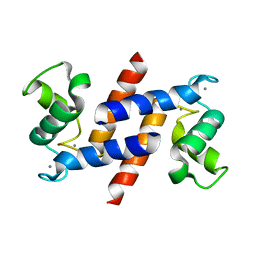
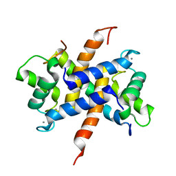
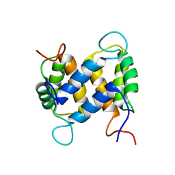
2WOS
 
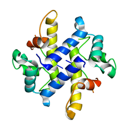
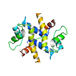
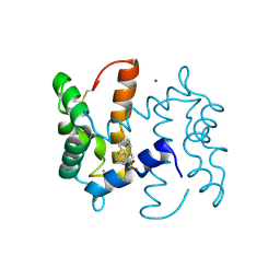 | | Structure of human S100A7 in complex with 2,6 ANS | | 分子名称: | 6-[(1E)-CYCLOHEXA-2,4-DIEN-1-YLIDENEAMINO]NAPHTHALENE-2-SULFONATE, CALCIUM ION, PROTEIN S100-A7, ... | | 著者 | Leon, R, Murray, J.I, Cragg, G, Farnell, B, Pace, T.C, Bohne, C, Boulanger, M.J, Hof, F. | | 登録日 | 2009-07-27 | | 公開日 | 2009-10-20 | | 最終更新日 | 2023-12-20 | | 実験手法 | X-RAY DIFFRACTION (1.7 Å) | | 主引用文献 | Identification and Characterization of Binding Sites on S100A7, a Participant in Cancer and Inflammation Pathways.
Biochemistry, 48, 2009
|
|
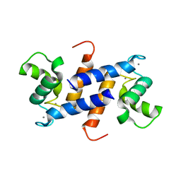
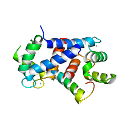
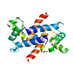
4GGF
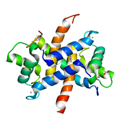
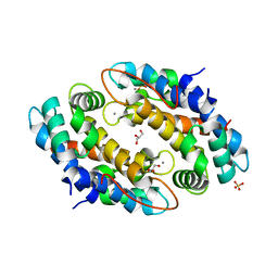 | | Crystal structure of Mn2+ bound calprotectin | | 分子名称: | CALCIUM ION, GLYCEROL, MANGANESE (II) ION, ... | | 著者 | Damo, S.M, Fritz, G. | | 登録日 | 2012-08-06 | | 公開日 | 2013-02-20 | | 最終更新日 | 2023-09-13 | | 実験手法 | X-RAY DIFFRACTION (1.6 Å) | | 主引用文献 | Molecular basis for manganese sequestration by calprotectin and roles in the innate immune response to invading bacterial pathogens.
Proc.Natl.Acad.Sci.USA, 110, 2013
|
|
3C1V
 | |
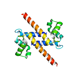
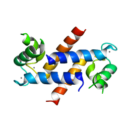
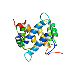
2WOR
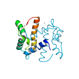 | | co-structure of S100A7 with 1,8 ANS | | 分子名称: | 8-ANILINO-1-NAPHTHALENE SULFONATE, CALCIUM ION, PROTEIN S100-A7, ... | | 著者 | Leon, R, Hof, F, Boulanger, M.J. | | 登録日 | 2009-07-27 | | 公開日 | 2009-10-20 | | 最終更新日 | 2023-12-20 | | 実験手法 | X-RAY DIFFRACTION (1.7 Å) | | 主引用文献 | Identification and Characterization of Binding Sites on S100A7, a Participant in Cancer and Inflammation Pathways.
Biochemistry, 48, 2009
|
|
3CGA
 | |
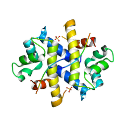
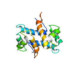
4HSZ
 | | Structure of truncated (delta8C) S100A4 | | 分子名称: | CALCIUM ION, Protein S100-A4 | | 著者 | Ramagopal, U.A, Dulyaninova, N.G, Kumar, P.R, Almo, S.C, Bresnick, A.R, New York Structural Genomics Research Consortium (NYSGRC) | | 登録日 | 2012-10-31 | | 公開日 | 2013-01-30 | | 最終更新日 | 2023-09-20 | | 実験手法 | X-RAY DIFFRACTION (2.25 Å) | | 主引用文献 | Structure of truncated (delta8C) S100A4
To be Published
|
|
2Y5I
 | |
4HRH
 | | Crystal Structure of p11-Annexin A2(N-terminal) Fusion Protein in Complex with SMARCA3 Peptide | | 分子名称: | Helicase-like transcription factor, Protein S100-A10, Annexin A2, ... | | 著者 | Gao, P, Patel, D.J. | | 登録日 | 2012-10-27 | | 公開日 | 2013-03-06 | | 最終更新日 | 2023-09-20 | | 実験手法 | X-RAY DIFFRACTION (3.001 Å) | | 主引用文献 | SMARCA3, a Chromatin-Remodeling Factor, Is Required for p11-Dependent Antidepressant Action.
Cell(Cambridge,Mass.), 152, 2013
|
|
4HRG
 | |
3ZWH
 | | Ca2+-bound S100A4 C3S, C81S, C86S and F45W mutant complexed with myosin IIA | | 分子名称: | ACETATE ION, AZIDE ION, CALCIUM ION, ... | | 著者 | Kiss, B, Duelli, A, Radnai, L, Kekesi, A.K, Katona, G, Nyitray, L. | | 登録日 | 2011-07-31 | | 公開日 | 2012-04-04 | | 最終更新日 | 2023-12-20 | | 実験手法 | X-RAY DIFFRACTION (1.94 Å) | | 主引用文献 | Crystal Structure of the S100A4-Nonmuscle Myosin Iia Tail Fragment Complex Reveals an Asymmetric Target Binding Mechanism.
Proc.Natl.Acad.Sci.USA, 109, 2012
|
|
4AQI
 | | Structure of human S100A15 bound to zinc and calcium | | 分子名称: | CALCIUM ION, CHLORIDE ION, PROTEIN S100-A7A, ... | | 著者 | Murray, J.I, Tonkin, M.L, Whiting, A.L, Peng, F, Farnell, B, Hof, F, Boulanger, M.J. | | 登録日 | 2012-04-17 | | 公開日 | 2012-10-17 | | 最終更新日 | 2023-12-20 | | 実験手法 | X-RAY DIFFRACTION (1.7 Å) | | 主引用文献 | Structural Characterization of S100A15 Reveals a Novel Zinc Coordination Site Among S100 Proteins and Altered Surface Chemistry with Functional Implications for Receptor Binding.
Bmc Struct.Biol., 12, 2012
|
|
4AQJ
 | | Structure of human S100A7 D24G bound to zinc and calcium | | 分子名称: | CALCIUM ION, CHLORIDE ION, PROTEIN S100-A7, ... | | 著者 | Murray, J.I, Tonkin, M.L, Whiting, A.L, Peng, F, Farnell, B, Hof, F, Boulanger, M.J. | | 登録日 | 2012-04-17 | | 公開日 | 2012-10-17 | | 実験手法 | X-RAY DIFFRACTION (1.6 Å) | | 主引用文献 | Structural Characterization of S100A15 Reveals a Novel Zinc Coordination Site Among S100 Proteins and Altered Surface Chemistry with Functional Implications for Receptor Binding.
Bmc Struct.Biol., 12, 2012
|
|
1CNP
 | | THE STRUCTURE OF CALCYCLIN REVEALS A NOVEL HOMODIMERIC FOLD FOR S100 CA2+-BINDING PROTEINS, NMR, 22 STRUCTURES | | 分子名称: | CALCYCLIN (RABBIT, APO) | | 著者 | Potts, B.C.M, Smith, J, Akke, M, Macke, T.J, Okazaki, K, Hidaka, H, Case, D.A, Chazin, W.J. | | 登録日 | 1995-08-31 | | 公開日 | 1996-10-14 | | 最終更新日 | 2022-02-16 | | 実験手法 | SOLUTION NMR | | 主引用文献 | The structure of calcyclin reveals a novel homodimeric fold for S100 Ca(2+)-binding proteins.
Nat.Struct.Biol., 2, 1995
|
|
4CFR
 | | Ca-bound S100A4 C3S, C81S, C86S and F45W mutant complexed with non- muscle myosin IIA | | 分子名称: | CALCIUM ION, MYOSIN-9, PROTEIN S100-A4 | | 著者 | Duelli, A, Kiss, B, Lundholm, I, Bodor, A, Radnai, L, Petoukhov, M, Svergun, D, Nyitray, L, Katona, G. | | 登録日 | 2013-11-19 | | 公開日 | 2014-05-07 | | 最終更新日 | 2023-12-20 | | 実験手法 | X-RAY DIFFRACTION (1.4 Å) | | 主引用文献 | The C-Terminal Random Coil Region Tunes the Ca2+-Binding Affinity of S100A4 Through Conformational Activation.
Plos One, 9, 2014
|
|
1E8A
 | | The three-dimensional structure of human S100A12 | | 分子名称: | CALCIUM ION, S100A12 | | 著者 | Moroz, O.V, Antson, A.A, Murshudov, G.N, Maitland, N.J, Dodson, G.G, Wilson, K.S, Skibshoj, I, Lukanidin, E.M, Bronstein, I.B. | | 登録日 | 2000-09-18 | | 公開日 | 2001-01-08 | | 最終更新日 | 2023-12-13 | | 実験手法 | X-RAY DIFFRACTION (1.95 Å) | | 主引用文献 | The Three-Dimensional Structure of Human S100A12
Acta Crystallogr.,Sect.D, 57, 2001
|
|
4CFQ
 | | Ca-bound truncated (delta13C) and C3S, C81S and C86S mutated S100A4 complexed with non-muscle myosin IIA | | 分子名称: | CALCIUM ION, MYOSIN-9, PROTEIN S100-A4 | | 著者 | Duelli, A, Kiss, B, Lundholm, I, Bodor, A, Radnai, L, Petoukhov, M, Svergun, D, Nyitray, L, Katona, G. | | 登録日 | 2013-11-19 | | 公開日 | 2014-05-07 | | 最終更新日 | 2023-12-20 | | 実験手法 | X-RAY DIFFRACTION (1.37 Å) | | 主引用文献 | The C-Terminal Random Coil Region Tunes the Ca2+-Binding Affinity of S100A4 Through Conformational Activation.
Plos One, 9, 2014
|
|
4DIR
 | |
4DUQ
 | |
4DRW
 | | Crystal Structure of the Ternary Complex between S100A10, an Annexin A2 N-terminal Peptide and an AHNAK Peptide | | 分子名称: | Neuroblast differentiation-associated protein AHNAK, Protein S100-A10/Annexin A2 chimeric protein | | 著者 | Rezvanpour, A, Lee, T.-W, Junop, M.S, Shaw, G.S. | | 登録日 | 2012-02-17 | | 公開日 | 2012-10-24 | | 最終更新日 | 2023-09-13 | | 実験手法 | X-RAY DIFFRACTION (3.5 Å) | | 主引用文献 | Structure of an asymmetric ternary protein complex provides insight for membrane interaction.
Structure, 20, 2012
|
|
3KO0
 | | Structure of the tfp-ca2+-bound activated form of the s100a4 Metastasis factor | | 分子名称: | 10-[3-(4-METHYL-PIPERAZIN-1-YL)-PROPYL]-2-TRIFLUOROMETHYL-10H-PHENOTHIAZINE, CALCIUM ION, Protein S100-A4 | | 著者 | Malashkevich, V.N, Dulyaninova, N.G, Knight, D, Almo, S.C, Bresnick, A.R. | | 登録日 | 2009-11-12 | | 公開日 | 2010-05-26 | | 最終更新日 | 2024-02-21 | | 実験手法 | X-RAY DIFFRACTION (2.3 Å) | | 主引用文献 | Phenothiazines inhibit S100A4 function by inducing protein oligomerization.
Proc.Natl.Acad.Sci.USA, 107, 2010
|
|
4ETO
 | | Structure of S100A4 in complex with non-muscle myosin-IIA peptide | | 分子名称: | CALCIUM ION, Myosin-9, Protein S100-A4 | | 著者 | Ramagopal, U.A, Dulyaninova, N.G, Kumar, P.R, Almo, S.C, Bresnick, A.R, New York Structural Genomics Research Consortium (NYSGRC), Atoms-to-Animals: The Immune Function Network (IFN) | | 登録日 | 2012-04-24 | | 公開日 | 2012-07-11 | | 最終更新日 | 2023-09-13 | | 実験手法 | X-RAY DIFFRACTION (1.54 Å) | | 主引用文献 | Structure of S100A4 with bound peptide P
To be Published
|
|
1YUR
 | | Solution structure of apo-S100A13 (minimized mean structure) | | 分子名称: | S100 calcium-binding protein A13 | | 著者 | Arnesano, F, Banci, L, Bertini, I, Fantoni, A, Tenori, L, Viezzoli, M.S, Structural Proteomics in Europe (SPINE) | | 登録日 | 2005-02-14 | | 公開日 | 2005-10-18 | | 最終更新日 | 2022-03-02 | | 実験手法 | SOLUTION NMR | | 主引用文献 | Structural Interplay between Calcium(II) and Copper(II) Binding to S100A13 Protein
Angew.Chem.Int.Ed.Engl., 44, 2005
|
|
1YUT
 | | Solution structure of Calcium-S100A13 (minimized mean structure) | | 分子名称: | CALCIUM ION, S100 calcium-binding protein A13 | | 著者 | Arnesano, F, Banci, L, Bertini, I, Fantoni, A, Tenori, L, Viezzoli, M.S, Structural Proteomics in Europe (SPINE) | | 登録日 | 2005-02-14 | | 公開日 | 2005-10-18 | | 最終更新日 | 2022-03-02 | | 実験手法 | SOLUTION NMR | | 主引用文献 | Structural Interplay between Calcium(II) and Copper(II) Binding to S100A13 Protein
Angew.Chem.Int.Ed.Engl., 44, 2005
|
|
1YUS
 | | Solution structure of apo-S100A13 | | 分子名称: | S100 calcium binding protein A13 | | 著者 | Arnesano, F, Banci, L, Bertini, I, Fantoni, A, Tenori, L, Viezzoli, M.S, Structural Proteomics in Europe (SPINE) | | 登録日 | 2005-02-14 | | 公開日 | 2005-10-18 | | 最終更新日 | 2022-03-02 | | 実験手法 | SOLUTION NMR | | 主引用文献 | Structural Interplay between Calcium(II) and Copper(II) Binding to S100A13 Protein
Angew.Chem.Int.Ed.Engl., 44, 2005
|
|
3M0W
 | | Structure of S100A4 with PCP | | 分子名称: | 1,4-DIETHYLENE DIOXIDE, 2-chloro-10-[3-(4-methylpiperazin-1-yl)propyl]-10H-phenothiazine, CALCIUM ION, ... | | 著者 | Ramagopal, U.A, Dulyaninova, N.G, Almo, S.C, Bresnick, A.R. | | 登録日 | 2010-03-03 | | 公開日 | 2010-05-12 | | 最終更新日 | 2023-09-06 | | 実験手法 | X-RAY DIFFRACTION (2.8 Å) | | 主引用文献 | Structure of S100A4 with PCP
To be published
|
|


