6MVK
 
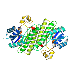
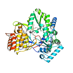 | | HCV NS5B 1b N316 bound to Compound 18 | | Descriptor: | (4-{(4S)-3-[5-cyclopropyl-2-(4-fluorophenyl)-3-(methylcarbamoyl)-1-benzofuran-6-yl]-2-oxo-1,3-oxazolidin-4-yl}-2-fluorophenyl)boronic acid, HCV Polymerase | | Authors: | Williams, S.P, Kahler, K, Price, D.J, Peat, A.J. | | Deposit date: | 2018-10-26 | | Release date: | 2019-09-04 | | Last modified: | 2024-03-13 | | Method: | X-RAY DIFFRACTION (2.3 Å) | | Cite: | Design of N-Benzoxaborole Benzofuran GSK8175-Optimization of Human Pharmacokinetics Inspired by Metabolites of a Failed Clinical HCV Inhibitor.
J.Med.Chem., 62, 2019
|
|
9RFE
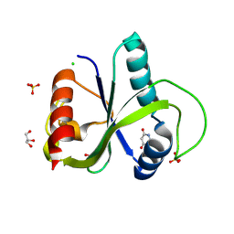
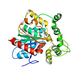
 | | Human ADP-ribosylhydrolase 3 (ARH3) in complex with an inhibitor | | Descriptor: | 2-[(2~{E})-2-[1-(4-aminophenyl)ethylidene]hydrazinyl]-6-methyl-1~{H}-pyrimidin-4-one, ACETIC ACID, ADP-ribosylhydrolase ARH3, ... | | Authors: | Paakkonen, J, Lehtio, L. | | Deposit date: | 2025-06-04 | | Release date: | 2025-09-24 | | Last modified: | 2025-10-29 | | Method: | X-RAY DIFFRACTION (1.85 Å) | | Cite: | Discovery and Structural Optimization of 2-Hydrazinopyrimidin-4-one Analogs Inhibiting Human ADP-Ribosylhydrolase ARH3.
Acs Chem.Biol., 20, 2025
|
|
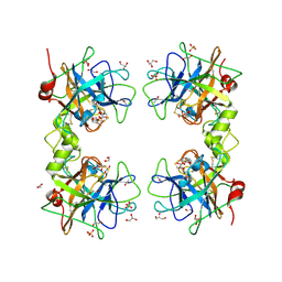
9QIU
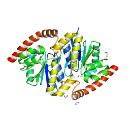
 | | Crystal structure of YTHDF2 in complex with compound 13 (AI-DF2-56) | | Descriptor: | 3-sulfanyl-1,2,4-triazin-5-ol, GLYCEROL, SULFATE ION, ... | | Authors: | Nai, F, Invernizzi, A, Caflisch, A. | | Deposit date: | 2025-03-17 | | Release date: | 2025-07-09 | | Last modified: | 2025-09-10 | | Method: | X-RAY DIFFRACTION (2.46 Å) | | Cite: | Discovery of YTHDF2 Ligands by Fragment-Based Design.
Acs Bio Med Chem Au, 5, 2025
|
|
9QEM
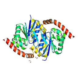
 | | Crystal structure of YTHDF2 in complex with compound 3 (AI-DF2-11) | | Descriptor: | 5-[[(3-methoxyphenyl)amino]methylidene]-2-sulfanylidene-1,3-diazinane-4,6-dione, CHLORIDE ION, GLYCEROL, ... | | Authors: | Nai, F, Invernizzi, A, Caflisch, A. | | Deposit date: | 2025-03-10 | | Release date: | 2025-07-09 | | Last modified: | 2025-09-10 | | Method: | X-RAY DIFFRACTION (2.26 Å) | | Cite: | Discovery of YTHDF2 Ligands by Fragment-Based Design.
Acs Bio Med Chem Au, 5, 2025
|
|
8TT5
 | | Pseudomonas fluorescens isocyanide hydratase pH=8.3 | | Descriptor: | 1,2-ETHANEDIOL, Isonitrile hydratase InhA | | Authors: | Wilson, M.A, Smith, N, Dasgupta, M, Dolamore, C. | | Deposit date: | 2023-08-12 | | Release date: | 2023-09-20 | | Last modified: | 2025-05-07 | | Method: | X-RAY DIFFRACTION (1.02 Å) | | Cite: | Changes in an enzyme ensemble during catalysis observed by high-resolution XFEL crystallography.
Sci Adv, 10, 2024
|
|
8TT2
 | | Pseudomonas fluorescens isocyanide hydratase pH=5.4 | | Descriptor: | 1,2-ETHANEDIOL, Isonitrile hydratase InhA | | Authors: | Wilson, M.A, Smith, N, Dasgupta, M, Dolamore, C. | | Deposit date: | 2023-08-12 | | Release date: | 2023-09-20 | | Last modified: | 2025-05-07 | | Method: | X-RAY DIFFRACTION (1.33 Å) | | Cite: | Changes in an enzyme ensemble during catalysis observed by high-resolution XFEL crystallography.
Sci Adv, 10, 2024
|
|
9RM4
 | | Crystal Structure of ZIKV NS2B-NS3 protease in complex with ASAP-0015373 | | Descriptor: | SODIUM ION, Serine protease subunit NS2B,Serine protease NS3, ~{N}-[3-chloranyl-5-[(2-methyl-1-oxidanyl-propan-2-yl)carbamoyl]phenyl]-5-[(dimethylamino)methyl]-1~{H}-pyrrole-2-carboxamide | | Authors: | Ni, X, Marples, P.G, Godoy, A.S, Koekemoer, L, Aschenbrenner, J.C, Balcomb, B.H, Fairhead, M, Lithgo, R.M, Lee, A, Kenton, N, Thompson, W, Tomlinson, C.W.E, Wild, C, Winokan, M, Williams, E.P, Fearon, D, von Delft, F. | | Deposit date: | 2025-06-17 | | Release date: | 2025-07-23 | | Method: | X-RAY DIFFRACTION (1.95 Å) | | Cite: | Crystal Structure of ZIKV NS2B-NS3 protease in complex with ASAP-0015373
To Be Published
|
|
8KDA
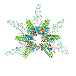 | | Cryo-EM structure of Hydrogenobacter thermophilus minimal protein-only RNase P (HARP) in complex with pre-tRNAs | | Descriptor: | Aquifex aeolicus pre-tRNAVal, MAGNESIUM ION, RNA-free ribonuclease P | | Authors: | Teramoto, T, Adachi, N, Yokogawa, T, Koyasu, T, Mayanagi, K, Nakamura, T, Senda, T, Kakuta, Y. | | Deposit date: | 2023-08-09 | | Release date: | 2024-08-14 | | Last modified: | 2025-07-16 | | Method: | ELECTRON MICROSCOPY (3.19 Å) | | Cite: | Structural basis of transfer RNA processing by bacterial minimal RNase P.
Nat Commun, 16, 2025
|
|
8TT4
 | | Pseudomonas fluorescens isocyanide hydratase pH=6.0 | | Descriptor: | 1,2-ETHANEDIOL, Isonitrile hydratase InhA | | Authors: | Wilson, M.A, Smith, N, Dasgupta, M, Dolamore, C. | | Deposit date: | 2023-08-12 | | Release date: | 2023-09-20 | | Last modified: | 2025-05-07 | | Method: | X-RAY DIFFRACTION (1.2 Å) | | Cite: | Changes in an enzyme ensemble during catalysis observed by high-resolution XFEL crystallography.
Sci Adv, 10, 2024
|
|
9SQ9
 | |
8C6Y
 | | PBP AccA from A. tumefaciens Bo542 in apoform 2 | | Descriptor: | 1,2-ETHANEDIOL, Agrocinopine utilization periplasmic binding protein AccA | | Authors: | Morera, S, Vigouroux, A, legrand, P. | | Deposit date: | 2023-01-12 | | Release date: | 2024-01-24 | | Last modified: | 2024-02-07 | | Method: | X-RAY DIFFRACTION (1.901 Å) | | Cite: | A highly conserved ligand-binding site for AccA transporters of antibiotic and quorum-sensing regulator in Agrobacterium leads to a different specificity.
Biochem.J., 481, 2024
|
|
9NXL
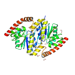
 | | Crystal structure of steroid aldehyde dehydrogenase (Sad) from Caldimonas tepidiphilia | | Descriptor: | (4S)-2-METHYL-2,4-PENTANEDIOL, 2-AMINO-2-HYDROXYMETHYL-PROPANE-1,3-DIOL, Steroid aldehyde dehydrogenase | | Authors: | Rolfe, N, Myskiw, D, Patton, M.T, Forrester, T.J.B, Kimber, M.S, Seah, S.Y.K. | | Deposit date: | 2025-03-25 | | Release date: | 2025-10-08 | | Method: | X-RAY DIFFRACTION (1.8 Å) | | Cite: | Sad from Proteobacteria is a Structurally Distinct ALDH3 Enzyme Specialized for the Oxidation of Steroidal Aldehydes.
Biochemistry, 64, 2025
|
|
9OCF
 | | 2.73A cryo-EM structure of the Measles Virus L-P in complex with ERdRp-0519 | | Descriptor: | 2-methyl-~{N}-[4-[(2~{S})-2-(2-morpholin-4-ylethyl)piperidin-1-yl]sulfonylphenyl]-5-(trifluoromethyl)pyrazole-3-carboxamide, Phosphoprotein, RNA-directed RNA polymerase L | | Authors: | Liu, B, Wang, D, Yang, G. | | Deposit date: | 2025-04-24 | | Release date: | 2025-09-03 | | Last modified: | 2025-10-29 | | Method: | ELECTRON MICROSCOPY (2.73 Å) | | Cite: | Structural basis of measles virus polymerase inhibition by nonnucleoside inhibitor ERDRP-0519.
Nat Commun, 16, 2025
|
|
9NIM
 | |
9RP9
 | |
9OXN
 | |
6Y1B
 | | X-ray structure of Lactobacillus brevis alcohol dehydrogenase mutant K32A_Q126K | | Descriptor: | 1,2-ETHANEDIOL, DI(HYDROXYETHYL)ETHER, MAGNESIUM ION, ... | | Authors: | Hermann, J, Bischoff, D, Janowski, R, Niessing, D, Grob, P, Hekmat, D, Weuster-Botz, D. | | Deposit date: | 2020-02-11 | | Release date: | 2020-02-19 | | Last modified: | 2024-01-24 | | Method: | X-RAY DIFFRACTION (1.4 Å) | | Cite: | Crystal Contact Engineering Enables Efficient Capture and Purification of an Oxidoreductase by Technical Crystallization.
Biotechnol J, 15, 2020
|
|
6N5G
 | | Crystal structure of an epoxide hydrolase from Trichoderma reesei in complex with inhibitor 2 | | Descriptor: | 4-[(quinolin-3-yl)methyl]-N-[4-(trifluoromethoxy)phenyl]piperidine-1-carboxamide, Epoxide hydrolase TrEH | | Authors: | Oliveira, G.S, Adriani, P.P, Ribeiro, J.A, Morisseau, C, Hammock, B.D, Dias, M.V, Chambergo, F.S. | | Deposit date: | 2018-11-21 | | Release date: | 2019-11-20 | | Last modified: | 2023-10-11 | | Method: | X-RAY DIFFRACTION (2.6 Å) | | Cite: | The molecular structure of an epoxide hydrolase from Trichoderma reesei in complex with urea or amide-based inhibitors.
Int. J. Biol. Macromol., 129, 2019
|
|
9QFV
 | | Human Tryptase beta-2 (hTPSB2) | | Descriptor: | 1,2-ETHANEDIOL, 2-(N-MORPHOLINO)-ETHANESULFONIC ACID, ACETATE ION, ... | | Authors: | Porta, A, Manelfi, C, Talarico, C, Beccari, A.R, Brindisi, M, Summa, V, Iaconis, D, Gobbi, M, Beeg, M. | | Deposit date: | 2025-03-12 | | Release date: | 2025-03-26 | | Last modified: | 2025-04-09 | | Method: | X-RAY DIFFRACTION (2.06 Å) | | Cite: | Integrating Surface Plasmon Resonance and Docking Analysis for Mechanistic Insights of Tryptase Inhibitors.
Molecules, 30, 2025
|
|
9QCY
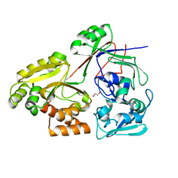
 | | Crystal structure of Rhizobium etli L-asparaginase ReAV S80A mutant | | Descriptor: | 1,2-ETHANEDIOL, CHLORIDE ION, DI(HYDROXYETHYL)ETHER, ... | | Authors: | Pokrywka, K, Grzechowiak, M, Loch, J.I, Ruszkowski, M, Gilski, M, Jaskolski, M. | | Deposit date: | 2025-03-05 | | Release date: | 2025-07-16 | | Last modified: | 2025-08-06 | | Method: | X-RAY DIFFRACTION (1.7 Å) | | Cite: | Probing the Active Site of Class 3 L-Asparaginase by Mutagenesis: Mutations of the Ser-Lys Tandems of ReAV.
Biomolecules, 15, 2025
|
|
9ON7
 | |
9PLN
 | | Locally-refined structure of alpha2a adrenergic receptor in complex with Go heterotrimer, scFv16, and N-(5-methylnaphthalen-1-yl)pyridin-4-amine (compound 4905) | | Descriptor: | Alpha-2A adrenergic receptor, N-(5-methylnaphthalen-1-yl)pyridin-4-amine | | Authors: | Srinivasan, K, Xu, X, Mailhot, O, Manglik, A, Shoichet, B. | | Deposit date: | 2025-07-15 | | Release date: | 2025-08-20 | | Method: | ELECTRON MICROSCOPY (2.8 Å) | | Cite: | Toward a Random Background for Ligand Optimization
To Be Published
|
|
9P4A
 | | E. coli Dihydropteroate Synthase in complex with pterin-based inhibitor | | Descriptor: | (2S)-2-(7-amino-4,5-dioxo-1,4,5,6-tetrahydropyrimido[4,5-c]pyridazin-3-yl)propanoic acid, Dihydropteroate synthase | | Authors: | Snoke, H.E, Reeve, S.M, Lee, R.E. | | Deposit date: | 2025-06-16 | | Release date: | 2025-11-05 | | Method: | X-RAY DIFFRACTION (3 Å) | | Cite: | Development of Pyrimido Pyridazine Analogs through Increased Whole Cell Target Engagement of the Dihydropteroate Synthase Pterin Binding Site in Gram-Negative Bacteria.
Acs Infect Dis., 2025
|
|
9OND
 | |
9QFY
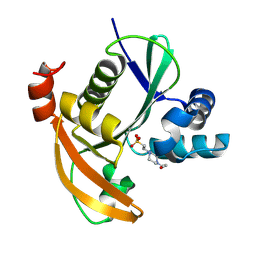
 | | Structure of CHIP E3 ubiquitin ligase TPR domain in complex with compound 9. | | Descriptor: | E3 ubiquitin-protein ligase CHIP, SULFATE ION, ~{N},~{N}-dimethyl-1-[(3~{S})-1-[[4,5,6,7-tetrakis(fluoranyl)-1~{H}-indol-3-yl]carbonyl]piperidin-3-yl]methanesulfonamide | | Authors: | Breed, J. | | Deposit date: | 2025-03-12 | | Release date: | 2025-08-27 | | Method: | X-RAY DIFFRACTION (1.064 Å) | | Cite: | Discovery of Small-Molecule Ligands for the E3 Ligase STUB1/CHIP from a DNA-Encoded Library Screen.
Acs Med.Chem.Lett., 16, 2025
|
|


