1B1E
 
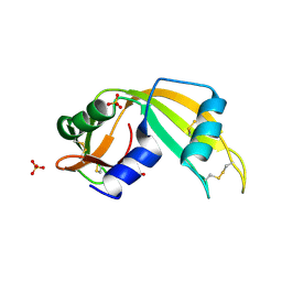
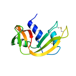
 | | CRYSTAL STRUCTURE OF HUMAN ANGIOGENIN VARIANT K40Q | | 分子名称: | CITRIC ACID, HYDROLASE ANGIOGENIN | | 著者 | Leonidas, D.D, Acharya, K.R. | | 登録日 | 1998-11-20 | | 公開日 | 1999-04-02 | | 最終更新日 | 2023-08-09 | | 実験手法 | X-RAY DIFFRACTION (2 Å) | | 主引用文献 | Refined crystal structures of native human angiogenin and two active site variants: implications for the unique functional properties of an enzyme involved in neovascularisation during tumour growth.
J.Mol.Biol., 285, 1999
|
|
1AQP
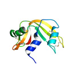
 | | RIBONUCLEASE A COPPER COMPLEX | | 分子名称: | COPPER (II) ION, RIBONUCLEASE A | | 著者 | Ramasubbu, N. | | 登録日 | 1997-07-31 | | 公開日 | 1998-05-27 | | 最終更新日 | 2023-08-02 | | 実験手法 | X-RAY DIFFRACTION (2 Å) | | 主引用文献 | Crystal structures of the copper and nickel complexes of RNase A: metal-induced interprotein interactions and identification of a novel copper binding motif.
Proc.Natl.Acad.Sci.USA, 94, 1997
|
|
1B1I
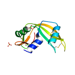
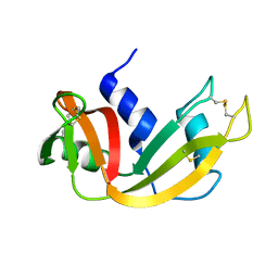
 | | CRYSTAL STRUCTURE OF HUMAN ANGIOGENIN | | 分子名称: | CITRIC ACID, HYDROLASE ANGIOGENIN | | 著者 | Leonidas, D.D, Acharya, K.R. | | 登録日 | 1998-11-20 | | 公開日 | 1999-04-02 | | 最終更新日 | 2023-08-09 | | 実験手法 | X-RAY DIFFRACTION (1.8 Å) | | 主引用文献 | Refined crystal structures of native human angiogenin and two active site variants: implications for the unique functional properties of an enzyme involved in neovascularisation during tumour growth.
J.Mol.Biol., 285, 1999
|
|
1BEL
 | | HYDROLASE PHOSPHORIC DIESTER, RNA | | 分子名称: | METHANOL, RIBONUCLEASE A, SULFATE ION | | 著者 | Dung, M.H, Bell, J.A. | | 登録日 | 1995-12-21 | | 公開日 | 1996-10-14 | | 最終更新日 | 2023-08-02 | | 実験手法 | X-RAY DIFFRACTION (1.6 Å) | | 主引用文献 | Structure of crystal form IX of bovine pancreatic ribonuclease A.
Acta Crystallogr.,Sect.D, 53, 1997
|
|
1BC4
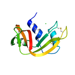
 | | THE SOLUTION STRUCTURE OF A CYTOTOXIC RIBONUCLEASE FROM THE OOCYTES OF RANA CATESBEIANA (BULLFROG), NMR, 15 STRUCTURES | | 分子名称: | RIBONUCLEASE | | 著者 | Chang, C.-F, Chen, C, Chen, Y.-C, Hom, K, Huang, R.-F, Huang, T. | | 登録日 | 1998-05-05 | | 公開日 | 1998-10-14 | | 最終更新日 | 2019-12-25 | | 実験手法 | SOLUTION NMR | | 主引用文献 | The solution structure of a cytotoxic ribonuclease from the oocytes of Rana catesbeiana (bullfrog).
J.Mol.Biol., 283, 1998
|
|
1BSR
 | |
1C0B
 | |
1C0C
 | |
1BZQ
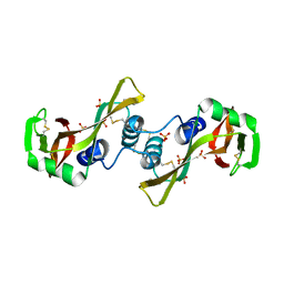
 | | COMPLEX OF A DROMEDARY SINGLE-DOMAIN VHH ANTIBODY FRAGMENT WITH RNASE A | | 分子名称: | PHOSPHATE ION, PROTEIN (ANTIBODY CAB-RN05), PROTEIN (RNASE A) | | 著者 | Decanniere, K, Desmyter, A, Gahroudhi, M, Lauwereys, M, Muyldermans, S, Wyns, L. | | 登録日 | 1998-11-03 | | 公開日 | 1998-11-11 | | 最終更新日 | 2023-08-09 | | 実験手法 | X-RAY DIFFRACTION (2.8 Å) | | 主引用文献 | A single-domain antibody fragment in complex with RNase A: non-canonical loop structures and nanomolar affinity using two CDR loops.
Structure Fold.Des., 7, 1999
|
|
1C8W
 | | THR45GLY VARIANT OF RIBONUCLEASE A | | 分子名称: | ACETATE ION, CHLORIDE ION, PROTEIN (Ribonuclease A) | | 著者 | Kelemen, B.R, Sweeney, R.T, Schultz, L.W, Raines, R.T. | | 登録日 | 1999-07-30 | | 公開日 | 2002-05-01 | | 最終更新日 | 2018-03-14 | | 実験手法 | X-RAY DIFFRACTION (1.8 Å) | | 主引用文献 | Excavating an active site: the nucleobase specificity of ribonuclease A.
Biochemistry, 39, 2000
|
|
1C9X
 | |
1C9V
 | |
1D5D
 | |
1D5E
 | |
1D5H
 | | Rnase s(f8a). mutant ribonucleasE S. | | 分子名称: | RNASE S, S PEPTIDE, SULFATE ION | | 著者 | Ratnaparkhi, G.S, Varadarajan, R. | | 登録日 | 1999-10-07 | | 公開日 | 1999-10-20 | | 最終更新日 | 2018-03-14 | | 実験手法 | X-RAY DIFFRACTION (2.25 Å) | | 主引用文献 | Thermodynamic and structural studies of cavity formation in proteins suggest that loss of packing interactions rather than the hydrophobic effect dominates the observed energetics.
Biochemistry, 39, 2000
|
|
1CJR
 | |
1CJQ
 | |
1E21
 | | Ribonuclease 1 des1-7 Crystal Structure at 1.9A | | 分子名称: | RIBONUCLEASE 1 | | 著者 | Pous, J, Mallorqui-Fernandez, G, Peracaula, R, Terzyan, S.S, Futami, J, Tada, H, Yamada, H, Seno, M, De Llorens, R, Gomis-Ruth, F.X, Coll, M. | | 登録日 | 2000-05-15 | | 公開日 | 2001-05-03 | | 最終更新日 | 2023-12-06 | | 実験手法 | X-RAY DIFFRACTION (1.9 Å) | | 主引用文献 | Three-Dimensional Crystal Structure of Human Rnase 1Dn7 at 1.9A Resolution
Acta Crystallogr.,Sect.D, 57, 2001
|
|
1DY5
 | | Deamidated derivative of bovine pancreatic ribonuclease | | 分子名称: | ACETATE ION, ISOPROPYL ALCOHOL, RIBONUCLEASE A, ... | | 著者 | Esposito, L, Vitagliano, L, Sica, F, Zagari, A, Mazzarella, L. | | 登録日 | 2000-01-27 | | 公開日 | 2000-03-28 | | 最終更新日 | 2023-12-06 | | 実験手法 | X-RAY DIFFRACTION (0.87 Å) | | 主引用文献 | The Ultrahigh Resolution Crystal Structure of Ribonuclease A Containing an Isoaspartyl Residue: Hydration and Sterochemical Analysis.
J.Mol.Biol., 297, 2000
|
|
1DZA
 | | 3-D structure of a HP-RNase | | 分子名称: | RIBONUCLEASE 1 | | 著者 | Pous, J, Canals, A, Terzyan, S.S, Guasch, A, Benito, A, Ribo, M, Vilanova, M, Coll, M. | | 登録日 | 2000-02-21 | | 公開日 | 2001-02-16 | | 最終更新日 | 2023-12-06 | | 実験手法 | X-RAY DIFFRACTION (1.65 Å) | | 主引用文献 | Three-Dimensional Structure of a Human Pancreatic Ribonuclease Variant, a Step Forward in the Design of Cytotoxic Ribonucleases
J.Mol.Biol., 303, 2000
|
|
1DYT
 | | X-ray crystal structure of ECP (RNase 3) at 1.75 A | | 分子名称: | CITRIC ACID, EOSINOPHIL CATIONIC PROTEIN, FE (III) ION | | 著者 | Mallorqui-Fernandez, G, Pous, J, Peracaula, R, Maeda, T, Tada, H, Yamada, H, Seno, M, De Llorens, R, Gomis-Rueth, F.X, Coll, M. | | 登録日 | 2000-02-08 | | 公開日 | 2001-02-08 | | 最終更新日 | 2024-05-01 | | 実験手法 | X-RAY DIFFRACTION (1.75 Å) | | 主引用文献 | Three-Dimensional Crystal Structure of Human Eosinophil Cationic Protein (Rnase 3) at 1.75 A Resolution.
J.Mol.Biol., 300, 2000
|
|
1EOS
 | | CRYSTAL STRUCTURE OF RIBONUCLEASE A COMPLEXED WITH URIDYLYL(2',5')GUANOSINE (PRODUCTIVE BINDING) | | 分子名称: | RIBONUCLEASE PANCREATIC, URIDYLYL-2'-5'-PHOSPHO-GUANOSINE | | 著者 | Vitagliano, L, Merlino, A, Zagari, A, Mazzarella, L. | | 登録日 | 2000-03-24 | | 公開日 | 2000-08-30 | | 最終更新日 | 2011-07-13 | | 実験手法 | X-RAY DIFFRACTION (2 Å) | | 主引用文献 | Productive and nonproductive binding to ribonuclease A: X-ray structure of two complexes with uridylyl(2',5')guanosine.
Protein Sci., 9, 2000
|
|
1EIE
 | |
1EID
 | |
1EIC
 | |


