Loading
PDBj
MenuPDBj@FacebookPDBj@TwitterPDBj@YouTubewwPDB FoundationwwPDB
RCSB PDBPDBeBMRBAdv. SearchSearch help
PDB: 3 results

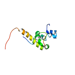
3KUS
DownloadVisualize
BU of 3kus by Molmil
Crystal structure of the MLLE domain of poly(A)-binding protein in complex with the binding region of Paip2
Descriptor: 4-(2-HYDROXYETHYL)-1-PIPERAZINE ETHANESULFONIC ACID, GLYCEROL, PAIP2 protein, ...
Authors:Kozlov, G, Gehring, K.
Deposit date:2009-11-27
Release date:2010-02-09
Last modified:2023-09-06
Method:X-RAY DIFFRACTION (1.4 Å)
Cite:Molecular Determinants of PAM2 Recognition by the MLLE Domain of Poly(A)-Binding Protein.
J.Mol.Biol., 397, 2010
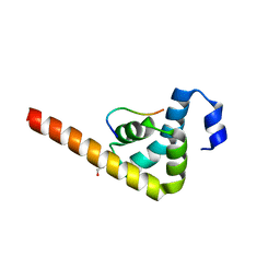
3KUT
DownloadVisualize
BU of 3kut by Molmil
Crystal structure of the MLLE domain of poly(A)-binding protein in complex with the binding region of Paip2
Descriptor: CHLORIDE ION, PAIP2 protein, Polyadenylate-binding protein 1
Authors:Kozlov, G, Gehring, K.
Deposit date:2009-11-27
Release date:2010-02-09
Last modified:2023-09-06
Method:X-RAY DIFFRACTION (1.5 Å)
Cite:Molecular Determinants of PAM2 Recognition by the MLLE Domain of Poly(A)-Binding Protein.
J.Mol.Biol., 397, 2010
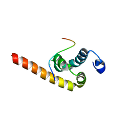
1JGN
DownloadVisualize
BU of 1jgn by Molmil
Solution structure of the C-terminal PABC domain of human poly(A)-binding protein in complex with the peptide from Paip2
Descriptor: polyadenylate-binding protein 1, polyadenylate-binding protein-interacting protein 2
Authors:Kozlov, G, Siddiqui, N, Coillet-Matillon, S, Ekiel, I, Gehring, K.
Deposit date:2001-06-26
Release date:2003-06-24
Last modified:2024-05-22
Method:SOLUTION NMR
Cite:Structural basis of ligand recognition by PABC, a highly specific peptide-binding domain found in poly(A)-binding protein and a HECT ubiquitin ligase
EMBO J., 23, 2004

223166

PDB entries from 2024-07-31

PDB statisticsPDBj update infoContact PDBjnumon