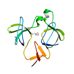
Loading
PDBj
MenuPDBj@FacebookPDBj@TwitterPDBj@YouTubewwPDB FoundationwwPDB
RCSB PDBPDBeBMRBAdv. SearchSearch help
PDB: 3 results

6AQB
DownloadVisualize
BU of 6aqb by Molmil
Structure of the SH3 domain of MLK3 bound to peptide generated from phage display
Descriptor: 1,2-ETHANEDIOL, Chimera protein of MLK3-SH3 and MIP (E.C.2.7.11.25), PHOSPHATE ION
Authors:Kall, S.K, Lavie, A.
Deposit date:2017-08-19
Release date:2018-07-04
Last modified:2024-03-13
Method:X-RAY DIFFRACTION (1.5 Å)
Cite:Identification of two distinct peptide-binding pockets in the SH3 domain of human mixed-lineage kinase 3.
J. Biol. Chem., 293, 2018
5K28
DownloadVisualize
BU of 5k28 by Molmil
Structure of the unbound SH3 domain of MLK3
Descriptor: Mitogen-activated protein kinase kinase kinase 11
Authors:Kall, S.K, Lavie, A.
Deposit date:2016-05-18
Release date:2017-12-13
Last modified:2023-09-27
Method:X-RAY DIFFRACTION (1.5 Å)
Cite:Identification of two distinct peptide-binding pockets in the SH3 domain of human mixed-lineage kinase 3.
J. Biol. Chem., 293, 2018
5K26
DownloadVisualize
BU of 5k26 by Molmil
Structure of the SH3 domain of MLK3 bound to peptide generated from phage display
Descriptor: 2-(N-MORPHOLINO)-ETHANESULFONIC ACID, Mitogen-activated protein kinase kinase kinase 11,Chimera protein of MLK3-SH3 and MIP
Authors:Kall, S.K, Lavie, A.
Deposit date:2016-05-18
Release date:2017-12-13
Last modified:2023-09-27
Method:X-RAY DIFFRACTION (1.2 Å)
Cite:Identification of two distinct peptide-binding pockets in the SH3 domain of human mixed-lineage kinase 3.
J. Biol. Chem., 293, 2018

221051

PDB entries from 2024-06-12

PDB statisticsPDBj update infoContact PDBjnumon