Loading
PDBj
MenuPDBj@FacebookPDBj@TwitterPDBj@YouTubewwPDB FoundationwwPDB
RCSB PDBPDBeBMRBAdv. SearchSearch help

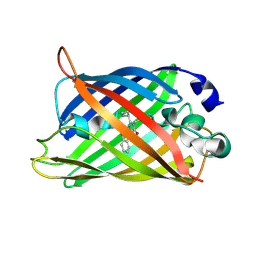
2EMO
DownloadVisualize
BU of 2emo by Molmil
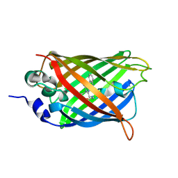
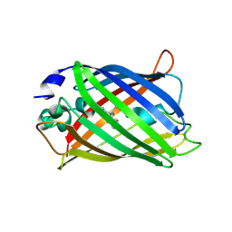
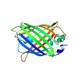
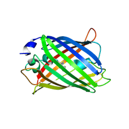
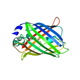
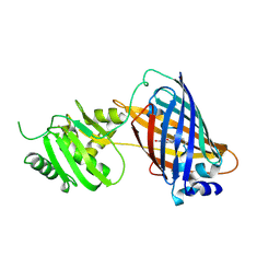
GREEN FLUORESCENT PROTEIN FROM AEQUOREA VICTORIA, MUTANT
Descriptor: GREEN FLUORESCENT PROTEIN
Authors:Palm, G, Zdanov, A, Wlodawer, A.
Deposit date:1997-03-31
Release date:1997-08-20
Last modified:2023-08-09
Method:X-RAY DIFFRACTION (2.6 Å)
Cite:The structural basis for spectral variations in green fluorescent protein.
Nat.Struct.Biol., 4, 1997
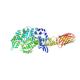
7K18
DownloadVisualize
BU of 7k18 by Molmil
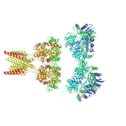
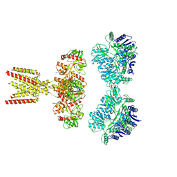
Cardiac Sodium channel with toxin bound
Descriptor: 2-acetamido-2-deoxy-beta-D-glucopyranose, 2-acetamido-2-deoxy-beta-D-glucopyranose-(1-4)-2-acetamido-2-deoxy-beta-D-glucopyranose, Alpha-like toxin Lqh3, ...
Authors:Jiang, D, Catterall, W.A.
Deposit date:2020-09-07
Release date:2021-01-20
Method:ELECTRON MICROSCOPY (3.3 Å)
Cite:Structural basis for voltage-sensor trapping of the cardiac sodium channel by a deathstalker scorpion toxin.
Nat Commun, 12, 2021
8T6L
DownloadVisualize
BU of 8t6l by Molmil
Cryo-EM structure of rat cardiac sodium channel NaV1.5 with batrachotoxin analog BTX-B
Descriptor: (1R)-1-[(5aR,7aR,9R,11aS,11bS,12R,13aR)-9,12-dihydroxy-2,11a-dimethyl-1,2,3,4,7a,8,9,10,11,11a,12,13-dodecahydro-7H-9,11b-epoxy-13a,5a-prop[1]enophenanthro[2,1-f][1,4]oxazepin-14-yl]ethyl benzoate, (3beta,14beta,17beta,25R)-3-[4-methoxy-3-(methoxymethyl)butoxy]spirost-5-en, 1-palmitoyl-2-oleoyl-sn-glycero-3-phosphocholine, ...
Authors:Tonggu, L, Wisedchaisri, G, Gamal El-Din, T.M, Zheng, N, Catterall, W.A.
Deposit date:2023-06-16
Release date:2024-03-06
Last modified:2024-03-27
Method:ELECTRON MICROSCOPY (3.3 Å)
Cite:Dual receptor-sites reveal the structural basis for hyperactivation of sodium channels by poison-dart toxin batrachotoxin.
Nat Commun, 15, 2024
8T18
DownloadVisualize
BU of 8t18 by Molmil
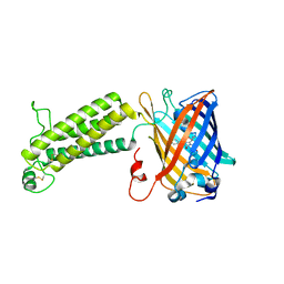
Cryo-EM structure of dodecameric hub domain of CaMKII beta
Descriptor: Venus-tagged CaMKII Alpha Association Domain
Authors:Chien, C.-T, Chiu, W, Khan, S.
Deposit date:2023-06-02
Release date:2024-06-12
Method:ELECTRON MICROSCOPY (2.6 Å)
Cite:Hub stability in the calcium calmodulin dependent protein kinase II
To Be Published
8T15
DownloadVisualize
BU of 8t15 by Molmil
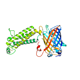
Cryo-EM structure of dodecameric hub domain of CaMKII alpha
Descriptor: Venus-tagged CaMKII Alpha Association Domain
Authors:Chien, C.-T, Chiu, W, Khan, S.
Deposit date:2023-06-01
Release date:2024-06-12
Method:ELECTRON MICROSCOPY (2.7 Å)
Cite:Hub stability in the calcium calmodulin dependent protein kinase II
To Be Published
8T17
DownloadVisualize
BU of 8t17 by Molmil
Cryo-EM structure of tetradecameric hub domain of CaMKII beta
Descriptor: Venus-tagged CaMKII Beta Association Domain
Authors:Chien, C.-T, Chiu, W, Khan, S.
Deposit date:2023-06-02
Release date:2024-06-12
Method:ELECTRON MICROSCOPY (2.6 Å)
Cite:Hub stability in the calcium calmodulin dependent protein kinase II
To Be Published
8T6K
DownloadVisualize
BU of 8t6k by Molmil
Cryo-EM structure of tetradecameric CaMKII beta holoenzyme T287A T306A T307A
Descriptor: Venus-tagged CaMKII Beta Holoenzyme mutant
Authors:Chien, C.-T, Chiu, W, Khan, S.
Deposit date:2023-06-16
Release date:2024-06-19
Method:ELECTRON MICROSCOPY (3 Å)
Cite:Hub stability in the calcium calmodulin dependent protein kinase II
To Be Published
8T6Q
DownloadVisualize
BU of 8t6q by Molmil
Cryo-EM structure of dodecameric CaMKII beta holoenzyme T287A T306A T307A
Descriptor: Venus-tagged CaMKII beta holoenzyme mutant
Authors:Chien, C.-T, Chiu, W, Khan, S.
Deposit date:2023-06-16
Release date:2024-06-19
Method:ELECTRON MICROSCOPY (3.5 Å)
Cite:Hub stability in the calcium calmodulin dependent protein kinase II
To Be Published
6UZ3
DownloadVisualize
BU of 6uz3 by Molmil
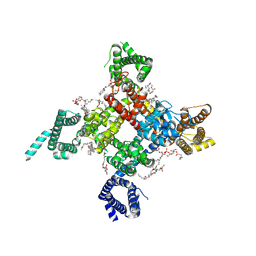
Cardiac sodium channel
Descriptor: (3beta,14beta,17beta,25R)-3-[4-methoxy-3-(methoxymethyl)butoxy]spirost-5-en, 2-acetamido-2-deoxy-beta-D-glucopyranose, 2-acetamido-2-deoxy-beta-D-glucopyranose-(1-4)-2-acetamido-2-deoxy-beta-D-glucopyranose, ...
Authors:Jiang, D, Shi, H, Tonggu, L, Lenaeus, M.J, Zheng, N, Catterall, W.A.
Deposit date:2019-11-14
Release date:2020-01-01
Last modified:2020-07-29
Method:ELECTRON MICROSCOPY (3.5 Å)
Cite:Structure of the Cardiac Sodium Channel.
Cell, 180, 2020
7KS0
DownloadVisualize
BU of 7ks0 by Molmil
GluK2/K5 with 6-Cyano-7-nitroquinoxaline-2,3-dione (CNQX)
Descriptor: Glutamate receptor ionotropic, kainate 2, kainate 5,Green fluorescent protein chimera
Authors:Khanra, N, Brown, P.M.G.E, Perozzo, A.M, Bowie, D, Meyerson, J.R.
Deposit date:2020-11-20
Release date:2021-03-24
Last modified:2021-07-07
Method:ELECTRON MICROSCOPY (5.3 Å)
Cite:Architecture and structural dynamics of the heteromeric GluK2/K5 kainate receptor.
Elife, 10, 2021
7KS3
DownloadVisualize
BU of 7ks3 by Molmil
GluK2/K5 with L-Glu
Descriptor: Glutamate receptor ionotropic, kainate 2, kainate 5,Green fluorescent protein chimera
Authors:Khanra, N, Brown, P.M.G.E, Perozzo, A.M, Bowie, D, Meyerson, J.R.
Deposit date:2020-11-20
Release date:2021-03-24
Last modified:2021-07-07
Method:ELECTRON MICROSCOPY (5.8 Å)
Cite:Architecture and structural dynamics of the heteromeric GluK2/K5 kainate receptor.
Elife, 10, 2021
6UZ0
DownloadVisualize
BU of 6uz0 by Molmil
Cardiac sodium channel with flecainide
Descriptor: (3beta,14beta,17beta,25R)-3-[4-methoxy-3-(methoxymethyl)butoxy]spirost-5-en, 2-acetamido-2-deoxy-beta-D-glucopyranose, 2-acetamido-2-deoxy-beta-D-glucopyranose-(1-4)-2-acetamido-2-deoxy-beta-D-glucopyranose, ...
Authors:Jiang, D, Shi, H, Tonggu, L, Lenaeus, M.J, Zheng, N, Catterall, W.A.
Deposit date:2019-11-14
Release date:2020-01-01
Last modified:2020-07-29
Method:ELECTRON MICROSCOPY (3.24 Å)
Cite:Structure of the Cardiac Sodium Channel.
Cell, 180, 2020
7KUY
DownloadVisualize
BU of 7kuy by Molmil
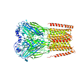
Cyro-EM structure of human Glycine Receptor alpha2-beta heteromer, strychnine bound state
Descriptor: 2-acetamido-2-deoxy-beta-D-glucopyranose, Glycine receptor subunit alpha-2, Glycine receptor subunit beta, ...
Authors:Yu, H, Wang, W.
Deposit date:2020-11-25
Release date:2021-09-08
Method:ELECTRON MICROSCOPY (3.6 Å)
Cite:Characterization of the subunit composition and structure of adult human glycine receptors
Neuron, 109, 2021
6WVI
DownloadVisualize
BU of 6wvi by Molmil
VKOR-like from Takifugu rubripes
Descriptor: (2R)-2,3-dihydroxypropyl (9Z)-octadec-9-enoate, Vitamin K epoxide reductase-like protein, termini restrained by green fluorescent protein
Authors:Liu, S, Sukumar, N, Li, W.
Deposit date:2020-05-06
Release date:2020-11-11
Last modified:2023-11-15
Method:X-RAY DIFFRACTION (2.4 Å)
Cite:Structural basis of antagonizing the vitamin K catalytic cycle for anticoagulation.
Science, 371, 2021
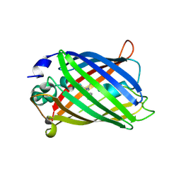
3V3D
DownloadVisualize
BU of 3v3d by Molmil
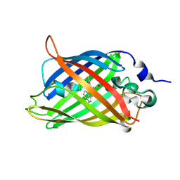
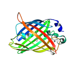
Crystal Structure of an eYFP single mutant
Descriptor: SULFATE ION, Yellow fluorescent protein
Authors:Ngan, N.B, Van Hecke, K, Van Meervelt, L.
Deposit date:2011-12-13
Release date:2012-12-26
Last modified:2023-12-06
Method:X-RAY DIFFRACTION (1.95 Å)
Cite:Improving the second-order nonlinear optical response of fluorescent proteins: the symmetry argument.
J.Am.Chem.Soc., 135, 2013
2P4M
DownloadVisualize
BU of 2p4m by Molmil
High pH structure of Rtms5 H146S variant
Descriptor: GFP-like non-fluorescent chromoprotein, IODIDE ION
Authors:Battad, J.M, Wilmann, P.G, Olsen, S, Byres, E, Smith, S.C, Dove, S.G, Turcic, K.N, Devenish, R.J, Rossjohn, J, Prescott, M.
Deposit date:2007-03-12
Release date:2007-04-03
Last modified:2023-11-15
Method:X-RAY DIFFRACTION (1.8 Å)
Cite:A structural basis for the pH-dependent increase in fluorescence efficiency of chromoproteins
J.Mol.Biol., 368, 2007
7SSX
DownloadVisualize
BU of 7ssx by Molmil
Structure of human Kv1.3
Descriptor: POTASSIUM ION, Potassium voltage-gated channel subfamily A member 3, Green fluorescent protein fusion
Authors:Meyerson, J.R, Selvakumar, P.
Deposit date:2021-11-11
Release date:2022-06-29
Last modified:2024-06-05
Method:ELECTRON MICROSCOPY (2.89 Å)
Cite:Structures of the T cell potassium channel Kv1.3 with immunoglobulin modulators.
Nat Commun, 13, 2022
7SSZ
DownloadVisualize
BU of 7ssz by Molmil
Structure of human Kv1.3 with A0194009G09 nanobodies
Descriptor: Nanobody A0194009G09, POTASSIUM ION, Potassium voltage-gated channel subfamily A member 3,Green fluorescent protein fusion
Authors:Meyerson, J.R, Selvakumar, P.
Deposit date:2021-11-11
Release date:2022-06-29
Last modified:2022-07-20
Method:ELECTRON MICROSCOPY (3.25 Å)
Cite:Structures of the T cell potassium channel Kv1.3 with immunoglobulin modulators.
Nat Commun, 13, 2022
7SSY
DownloadVisualize
BU of 7ssy by Molmil
Structure of human Kv1.3 (alternate conformation)
Descriptor: POTASSIUM ION, Potassium voltage-gated channel subfamily A member 3,Green fluorescent protein fusion
Authors:Meyerson, J.R, Selvakumar, P.
Deposit date:2021-11-11
Release date:2022-06-29
Last modified:2024-06-05
Method:ELECTRON MICROSCOPY (2.89 Å)
Cite:Structures of the T cell potassium channel Kv1.3 with immunoglobulin modulators.
Nat Commun, 13, 2022
7SSV
DownloadVisualize
BU of 7ssv by Molmil
Structure of human Kv1.3 with Fab-ShK fusion
Descriptor: Fab-ShK fusion, heavy chain, light chain, ...
Authors:Meyerson, J.R, Selvakumar, P, Smider, V, Huang, R.
Deposit date:2021-11-11
Release date:2022-06-29
Last modified:2022-07-20
Method:ELECTRON MICROSCOPY (3.39 Å)
Cite:Structures of the T cell potassium channel Kv1.3 with immunoglobulin modulators.
Nat Commun, 13, 2022
8F6P
DownloadVisualize
BU of 8f6p by Molmil
Rat Cardiac Sodium Channel with Ranolazine Bound
Descriptor: (R)-ranolazine, 2-acetamido-2-deoxy-beta-D-glucopyranose, 2-acetamido-2-deoxy-beta-D-glucopyranose-(1-4)-2-acetamido-2-deoxy-beta-D-glucopyranose, ...
Authors:Lenaeus, M.J, Tonggu, L.
Deposit date:2022-11-16
Release date:2023-04-12
Method:ELECTRON MICROSCOPY (3.2 Å)
Cite:Structural Basis for Inhibition of the Cardiac Sodium Channel by the Atypical Antiarrhythmic Drug Ranolazine
To Be Published
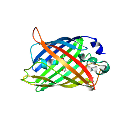
1KYP
DownloadVisualize
BU of 1kyp by Molmil
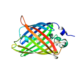
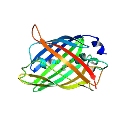
Crystal Structure of an Apo Green Fluorescent Protein Zn Biosensor
Descriptor: Green Fluorescent Protein, MAGNESIUM ION
Authors:Barondeau, D.P, Kassmann, C.J, Tainer, J.A, Getzoff, E.D.
Deposit date:2002-02-05
Release date:2002-04-10
Last modified:2023-11-15
Method:X-RAY DIFFRACTION (1.35 Å)
Cite:Structural chemistry of a green fluorescent protein Zn biosensor.
J.Am.Chem.Soc., 124, 2002
1KYS
DownloadVisualize
BU of 1kys by Molmil
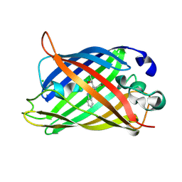
Crystal Structure of a Zn-bound Green Fluorescent Protein Biosensor
Descriptor: Green Fluorescent Protein, ZINC ION
Authors:Barondeau, D.P, Kassmann, C.J, Tainer, J.A, Getzoff, E.D.
Deposit date:2002-02-05
Release date:2002-04-10
Last modified:2023-11-15
Method:X-RAY DIFFRACTION (1.44 Å)
Cite:Structural chemistry of a green fluorescent protein Zn biosensor.
J.Am.Chem.Soc., 124, 2002
1KYR
DownloadVisualize
BU of 1kyr by Molmil
Crystal Structure of a Cu-bound Green Fluorescent Protein Zn Biosensor
Descriptor: COPPER (II) ION, Green Fluorescent Protein, MAGNESIUM ION
Authors:Barondeau, D.P, Kassmann, C.J, Tainer, J.A, Getzoff, E.D.
Deposit date:2002-02-05
Release date:2002-04-10
Last modified:2023-11-15
Method:X-RAY DIFFRACTION (1.5 Å)
Cite:Structural chemistry of a green fluorescent protein Zn biosensor.
J.Am.Chem.Soc., 124, 2002
1MOU
DownloadVisualize
BU of 1mou by Molmil
Crystal structure of Coral pigment
Descriptor: GFP-like non-fluorescent chromoprotein, IODIDE ION
Authors:Prescott, M, Ling, M, Beddoe, T, Oakley, A.J, Dove, S, Hoegh-Guldberg, O, Devenish, R.J, Rossjohn, J.
Deposit date:2002-09-10
Release date:2003-04-08
Last modified:2023-11-15
Method:X-RAY DIFFRACTION (2.2 Å)
Cite:The 2.2 a crystal structure of a pocilloporin pigment reveals a nonplanar chromophore conformation.
Structure, 11, 2003

222036

數據於2024-07-03公開中

PDB statisticsPDBj update infoContact PDBjnumon