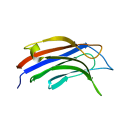
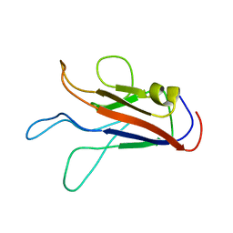
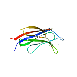
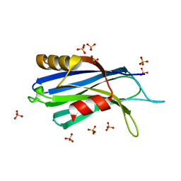
6NYC
 
 | | Munc13-1 C2B-domain, calcium free | | Descriptor: | 2-[3-(2-HYDROXY-1,1-DIHYDROXYMETHYL-ETHYLAMINO)-PROPYLAMINO]-2-HYDROXYMETHYL-PROPANE-1,3-DIOL, CHLORIDE ION, Munc13-1 | | Authors: | Tomchick, D.R, Rizo, J, Machius, M, Lu, J. | | Deposit date: | 2019-02-11 | | Release date: | 2019-02-20 | | Last modified: | 2023-10-11 | | Method: | X-RAY DIFFRACTION (1.893 Å) | | Cite: | Munc13 C2B domain is an activity-dependent Ca2+ regulator of synaptic exocytosis.
Nat. Struct. Mol. Biol., 17, 2010
|
|
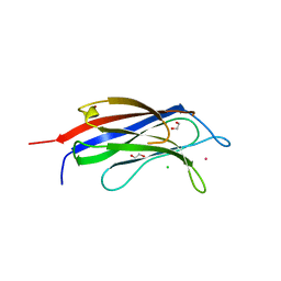
9BKY
 | |
9BKW
 | |
5A52
 | |
5A4X
 | |
5A50
 | |
7K6B
 | |
7KRB
 | |
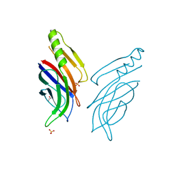
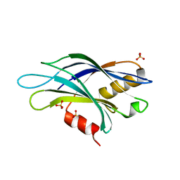
3RDJ
 | | Rat PKC C2 domain Apo | | Descriptor: | Protein kinase C alpha type | | Authors: | Li, P. | | Deposit date: | 2011-04-01 | | Release date: | 2011-09-07 | | Last modified: | 2024-02-21 | | Method: | X-RAY DIFFRACTION (1.9 Å) | | Cite: | Pb2+ as modulator of protein-membrane interactions.
J.Am.Chem.Soc., 133, 2011
|
|
7JOF
 | | Calcium-bound C2A Domain from Human Dysferlin | | Descriptor: | CALCIUM ION, Isoform 6 of Dysferlin | | Authors: | Tadayon, R, Wang, Y, Santamaria, L, Mercier, P, Forristal, C, Shaw, G.S. | | Deposit date: | 2020-08-06 | | Release date: | 2021-06-16 | | Last modified: | 2023-10-18 | | Method: | X-RAY DIFFRACTION (2 Å) | | Cite: | Calcium binds and rigidifies the dysferlin C2A domain in a tightly coupled manner.
Biochem.J., 478, 2021
|
|
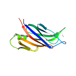
3HN8
 | | Crystal structure of synaptotagmin | | Descriptor: | CALCIUM ION, Synaptotagmin-3, ZINC ION | | Authors: | Strop, P, Vrljic, M, Ernst, J, Brunger, A.T. | | Deposit date: | 2009-05-30 | | Release date: | 2010-02-23 | | Last modified: | 2017-11-01 | | Method: | X-RAY DIFFRACTION (3.5 Å) | | Cite: | Molecular mechanism of the synaptotagmin-SNARE interaction in Ca2+-triggered vesicle fusion.
Nat.Struct.Mol.Biol., 17, 2010
|
|
3L9B
 | | Crystal Structure of Rat Otoferlin C2A | | Descriptor: | MAGNESIUM ION, Otoferlin | | Authors: | Helfmann, S, Neumann, P. | | Deposit date: | 2010-01-04 | | Release date: | 2011-01-19 | | Last modified: | 2023-11-01 | | Method: | X-RAY DIFFRACTION (1.95 Å) | | Cite: | The crystal structure of the C2A domain of otoferlin reveals an unconventional top loop region.
J.Mol.Biol., 406, 2011
|
|
3RPB
 | | THE C2B-DOMAIN OF RABPHILIN: STRUCTURAL VARIATIONS IN A JANUS-FACED DOMAIN | | Descriptor: | RABPHILIN 3-A | | Authors: | Ubach, J, Garcia, J, Nittler, M.P, Sudhof, T.C, Rizo, J. | | Deposit date: | 1999-04-19 | | Release date: | 1999-12-23 | | Last modified: | 2024-05-22 | | Method: | SOLUTION NMR | | Cite: | Structure of the Janus-faced C2B domain of rabphilin.
Nat.Cell Biol., 1, 1999
|
|
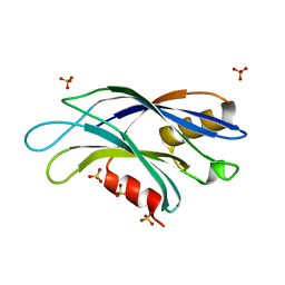
3TWY
 | | RAT PKC C2 DOMAIN BOUND TO PB | | Descriptor: | LEAD (II) ION, Protein kinase C alpha type, SULFATE ION | | Authors: | Li, P. | | Deposit date: | 2011-09-22 | | Release date: | 2011-11-02 | | Last modified: | 2024-02-28 | | Method: | X-RAY DIFFRACTION (1.5 Å) | | Cite: | Pb2+ as Modulator of Protein-Membrane Interactions.
J.Am.Chem.Soc., 133, 2011
|
|
7A1R
 | |
7ATP
 | |
7AS6
 | | 2.0 angstrom structure of plant Extended Synaptotagmin 1, C2A domain | | Descriptor: | 1,2-ETHANEDIOL, CADMIUM ION, CHLORIDE ION, ... | | Authors: | Benavente, J.L, Albert, A. | | Deposit date: | 2020-10-27 | | Release date: | 2021-08-18 | | Last modified: | 2024-01-31 | | Method: | X-RAY DIFFRACTION (2 Å) | | Cite: | The structure and flexibility analysis of the Arabidopsis synaptotagmin 1 reveal the basis of its regulation at membrane contact sites.
Life Sci Alliance, 4, 2021
|
|
6TZ3
 | | Crystal Structure of Human Synaptotagmin 1 C2B without Ca2+ | | Descriptor: | SULFATE ION, Synaptotagmin-1 | | Authors: | Dominguez, M.J, Karmakar, S, Meyer, A.G, Sutton, R.B. | | Deposit date: | 2019-08-09 | | Release date: | 2020-05-13 | | Last modified: | 2023-10-11 | | Method: | X-RAY DIFFRACTION (1.17 Å) | | Cite: | Molecular Basis for Synaptotagmin-1-Associated Neurodevelopmental Disorder.
Neuron, 107, 2020
|
|
6U41
 | | 1.7 angstrom structure of a pathogenic human Syt 1 C2B (D304G) | | Descriptor: | SULFATE ION, Synaptotagmin-1 | | Authors: | Dominguez, M.J, Bradberry, M.M, Chapman, E.R, Sutton, R.B. | | Deposit date: | 2019-08-22 | | Release date: | 2020-05-13 | | Last modified: | 2023-10-11 | | Method: | X-RAY DIFFRACTION (1.7 Å) | | Cite: | Molecular Basis for Synaptotagmin-1-Associated Neurodevelopmental Disorder.
Neuron, 107, 2020
|
|
6U4U
 | | 1.3 A structure of a pathogenic human Syt 1 C2B (I368T) | | Descriptor: | SULFATE ION, Synaptotagmin-1 | | Authors: | Dominguez, M.J, Bradberry, M.M, Chapman, E.R, Sutton, R.B. | | Deposit date: | 2019-08-26 | | Release date: | 2020-05-13 | | Last modified: | 2023-10-11 | | Method: | X-RAY DIFFRACTION (1.3 Å) | | Cite: | Molecular Basis for Synaptotagmin-1-Associated Neurodevelopmental Disorder.
Neuron, 107, 2020
|
|
1WFJ
 | |
1WFM
 | |
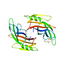
3N5A
 | | Synaptotagmin-7, C2B-domain, calcium bound | | Descriptor: | CALCIUM ION, Synaptotagmin-7 | | Authors: | Tomchick, D.R, Rizo, J, Craig, T.K. | | Deposit date: | 2010-05-24 | | Release date: | 2010-09-29 | | Last modified: | 2023-09-06 | | Method: | X-RAY DIFFRACTION (1.441 Å) | | Cite: | Structural and mutational analysis of functional differentiation between synaptotagmins-1 and -7.
Plos One, 5, 2010
|
|
6U4W
 | | 1.4 A structure of a pathogenic human Syt 1 C2B (D366E) | | Descriptor: | SULFATE ION, Synaptotagmin-1 | | Authors: | Dominguez, M.J, Bradberry, M.M, Chapman, E.R, Sutton, R.B. | | Deposit date: | 2019-08-26 | | Release date: | 2020-05-13 | | Last modified: | 2023-10-11 | | Method: | X-RAY DIFFRACTION (1.4 Å) | | Cite: | Molecular Basis for Synaptotagmin-1-Associated Neurodevelopmental Disorder.
Neuron, 107, 2020
|
|
2NCE
 | |


